LAST MEDICALLY REVIEWED:
SEPTEMBER 2026 — Dr. Shaileshkumar Garge
Citi Vascular Hospital, KPHB Colony, Road No. 1, Hyderabad, Telangana 500072
QUICK ANSWER
What Is Pelvic Venous Embolisation for Erectile Dysfunction — and Does It Work?
Pelvic venous embolisation is a minimally invasive endovascular procedure for carefully selected men with erectile dysfunction caused by venous leak (veno-occlusive dysfunction) — confirmed by pharmacological penile Doppler and CT cavernosography. Abnormal venous drainage pathways are mapped and selectively closed through catheter-based embolisation. The most recent multicentre registry (175 patients, 2025) reports 99.5% technical success and 59.4% meeting predefined clinical improvement at approximately 9 months. It is not appropriate for every man who has difficulty maintaining an erection. Proper diagnosis comes first. Dr. Garge FRCR (UK) | Citi Vascular Centre, KPHB, Hyderabad | +91-73375 83901.
'I can get an erection but I cannot keep it' is one of the most common complaints men bring to erectile dysfunction consultations. The frustration is real, the impact on relationships and self-esteem is significant, and the desire for a solution is understandable. But this symptom — an erection that initiates but rapidly softens — is not diagnostic of any single cause. Arterial disease, diabetes, medication side effects, Peyronie's disease, neurological factors, and psychological overlay can all produce the same complaint. One specific vascular mechanism — venous leak, more accurately called veno-occlusive dysfunction — is the target of pelvic venous embolisation. But it must be demonstrated, not assumed.
Pelvic venous embolisation for erectile dysfunction is one of the most clinically sophisticated endovascular procedures in vascular medicine — because it requires not only catheter skill but a precise understanding of penile venous anatomy, the ability to interpret CT cavernosography, and the technical judgement to selectively occlude abnormal drainage pathways without causing non-target embolisation. Recent publication of multicentre registry data with over 175 patients, and a 2026 report of more than 1,000 treatments from a dedicated endovascular ED programme, gives this procedure a growing and specific evidence base that informs what patients can realistically expect. This page reviews everything a patient considering this procedure should understand — including the honest complication data, the realistic success rates, and who is and is not a good candidate.
Book your Appointment here:
|| Appoinment: || Call +91-7032508468 || Whatsapp:+91-7337583901 || Citi Vascular Hospital, KPHB Colony, Hyderabad.||
|
Feature |
Detail |
|
Procedure Type |
Minimally invasive endovascular catheter procedure — no open surgery, no surgical incision on the penis or perineum |
|
Target |
Abnormal venous drainage from the corpus cavernosum — deep dorsal vein, cavernous veins, pudendal veins, periprostatic venous plexus |
|
Indication |
Confirmed venogenic / veno-occlusive erectile dysfunction — not every man who 'cannot maintain an erection' qualifies. Doppler + CT cavernosography required. |
|
Access Route |
Anterograde access via deep dorsal penile vein (most published series) | Retrograde pelvic venous access | Technique depends on anatomy and operator |
|
Key Diagnostic Investigation |
Pharmacological penile Doppler (EDV > 5 cm/s + RI < 0.75) PLUS CT cavernosography to map venous anatomy and confirm treatable pathways |
|
2025 Multicentre Registry Data (175 patients) |
Technical success: 99.5% | Clinical improvement (predefined endpoint): 59.4% at ~9 months | Recurrence/persistence: 10.2% | PE (non-target embolisation): 3 patients — no long-term consequences reported |
|
Anaesthesia |
Local anaesthesia + IV sedation — general anaesthesia not required in most cases |
|
Hospital Stay |
Same-day or overnight discharge in most cases |
A normal erection is a carefully orchestrated vascular event requiring two simultaneous mechanisms to work: arterial inflow increasing under the stimulus of sexual arousal, and venous occlusion preventing that blood from draining away before adequate intracavernous pressure builds. It is this second mechanism — the veno-occlusive mechanism — that fails in venogenic erectile dysfunction.
During erection, as the corpus cavernosum expands with incoming arterial blood, the subtunical venular plexus — the network of small veins immediately beneath the tunica albuginea — is compressed between the expanding cavernous tissue and the rigid tunica. This compression physically pinches the emissary veins that would otherwise drain blood from the penis, trapping the blood within the corpus cavernosum and maintaining the high intracavernous pressure (60–100 mmHg) required for rigidity. When this mechanism fails — because the tunica albuginea is structurally damaged, the cavernous smooth muscle cannot relax adequately, or the subtunical venous plexus is abnormally large or incompetent — blood continues to drain despite ongoing arterial inflow.
Normal Erection
Arterial inflow ↑↓Corpus cavernosum expands↓Subtunical veins compressed against tunica albuginea↓Venous outflow blocked↓Intracavernous pressure maintained at 60–100 mmHg↓Firm, sustained erection
Veno-Occlusive Dysfunction
Arterial inflow ↑ (may be adequate)↓Corpus cavernosum expands↓Subtunical veins NOT adequately compressed — remain open↓Excessive venous outflow continues↓Intracavernous pressure cannot be maintained↓Erection initiates but rapidly softens
Critical clinical point — symptoms alone do not diagnose venous leak: 'I can get an erection but cannot keep it' is a symptom, not a diagnosis. The same pattern occurs in arterial insufficiency, diabetes, medication side effects (antihypertensives, antidepressants, antiandrogens), Peyronie's disease, neurological conditions, and performance anxiety. A proper diagnostic assessment — pharmacological penile Doppler plus CT cavernosography — is essential before any consideration of venous embolisation. At Citi Vascular Centre, KPHB, the diagnostic assessment is the first and most important step. Call +91-73375 83901.
|
Cause |
How It Produces Venous Leak |
|
Peyronie's Disease |
Fibrous plaque deposition in the tunica albuginea prevents normal circumferential expansion — the tunica cannot adequately compress the subtunical venous plexus during tumescence. Most common identifiable structural cause of venous leak in younger men. The Peyronie's itself requires assessment alongside venous embolisation planning. |
|
Post-Radical Prostatectomy |
Autonomic denervation of cavernous smooth muscle — without adequate nitric oxide-mediated relaxation, the smooth muscle cannot fully dilate, reducing the mechanical compression of subtunical veins. Venous leak in this context has a significant neurogenic component alongside the venous mechanism, which limits the response to embolisation. |
|
Diabetes mellitus |
Chronic hyperglycaemia causes glycation and fibrosis of cavernous smooth muscle, reducing its relaxation capacity. Autonomic neuropathy further impairs the nitric oxide-mediated relaxation that drives full tumescence. Both mechanisms reduce veno-occlusion. |
|
Ageing-related cavernous degeneration |
Progressive replacement of cavernous smooth muscle by connective tissue and fibrosis — reducing elastic compliance of the corpus cavernosum and imparing veno-occlusion capacity. Common in men over 60. |
|
Idiopathic / Congenitally large venous channels |
Some younger men present with venous leak without identifiable structural disease — possibly from congenitally large or aberrant emissary veins, subtunical venous plexus abnormalities, or large accessory draining veins. This subgroup has the most favourable response to venous embolisation. |
|
Chronic perineal compression (cyclists) |
Prolonged narrow saddle cycling causes perineal compression of the pudendal neurovascular bundle — affecting both arterial inflow and venous drainage. May produce both arterial and venous ED components. |
|
Pelvic trauma |
Disruption of the tunica albuginea or emissary veins from penile or perineal trauma can create structural venous leak. |
The diagnostic pathway for venous leak erectile dysfunction is sequential and evidence-based. Each step builds on the last, and the decision to proceed to pelvic venous embolisation is only appropriate after all three levels of assessment have been completed and have confirmed venous leak as the treatable vascular mechanism.
|
1 |
Step 1 — Clinical History, IIEF Score, and Hormonal Assessment The IIEF-15 (International Index of Erectile Function) documents baseline erectile function across 5 domains. Clinical history establishes onset pattern, morning erection status, PDE5 inhibitor response, cardiovascular risk factors, medications (antihypertensives, antidepressants, antiandrogens), previous pelvic surgery or trauma, Peyronie's disease, diabetes, and psychological or relationship context. Testosterone (total and free), LH, prolactin, and TSH are measured — hormonal deficiency must be excluded and corrected before vascular testing. |
|
2 |
Step 2 — Pharmacological Penile Doppler Ultrasound After intracavernosal injection of prostaglandin E1 (PGE1, 10–20 micrograms), the cavernous arteries are assessed with duplex Doppler at 5-minute intervals. PSV, EDV, and RI are measured. Venous leak is suspected when: EDV remains > 5 cm/s at peak tumescence — indicating persistent forward venous flow that should have ceased as the veno-occlusive mechanism engages; AND RI < 0.75 — indicating inadequate resistance from venous closure. PSV should be > 25 cm/s, confirming adequate arterial inflow and identifying the venous mechanism as the primary problem. If PSV is also low, mixed arterial and venous disease is present — the arterial component is addressed first. Doppler alone is not sufficient to confirm venous leak in isolation — it must be complemented by anatomical venous imaging. |
|
3 |
Step 3 — CT Cavernosography or Venography — Mapping the Abnormal Drainage CT cavernosography is the key anatomical investigation for venous leak. A vasoactive agent is injected intracavernosally to produce maximal tumescence, followed by contrast injection directly into the corpus cavernosum. CT images map the entire venous drainage tree under active tumescent conditions — identifying the deep dorsal penile vein, cavernous veins, pudendal venous plexus, periprostatic veins, and any accessory draining channels. This imaging identifies: which veins are the dominant abnormal drainage pathways; whether the anatomy is suitable for catheter-based embolisation; and which access route (anterograde deep dorsal vein vs retrograde pelvic venous) is most appropriate. Published studies specifically emphasise CT cavernosography as the imaging modality required for appropriate patient selection and treatment planning in venous embolisation for ED. |
Careful patient selection is the single most important determinant of outcome in pelvic venous embolisation for erectile dysfunction. The procedure should not be offered to every man with difficulty maintaining an erection — the diagnostic pathway in Section 5 must be completed, the venous leak must be confirmed objectively, and the anatomy must be suitable for catheter-based embolisation.
|
Favourable for Venous Embolisation EDV > 5 cm/s + RI < 0.75 on pharmacological Doppler | PSV > 25 cm/s (adequate arterial inflow) | CT cavernosography confirms anatomically treatable venous drainage pathways | Failed adequate PDE5 inhibitor trial (two agents at maximum dose) | Erection initiates but cannot be maintained — classic veno-occlusive symptom | Idiopathic venous leak in younger men without Peyronie's or prostatectomy | Realistic expectations about improvement probability (59.4% clinical improvement in 2025 registry) |
Less Favourable — Consider Carefully Severe Peyronie's disease with significant structural damage — treat Peyronie's first | Post-radical prostatectomy — neurogenic component limits embolisation response | Severe cavernous smooth muscle atrophy (MRI or biopsy) — structural fibrosis is not reversible by venous embolisation | Mixed arterial + venous ED — treat arterial component first with angioplasty; reassess venous on Doppler | Predominantly psychogenic ED — embolisation does not treat psychological mechanism | Coagulopathy not correctable |
Important — mixed arterial and venous ED: When pharmacological Doppler shows both low PSV (arterial insufficiency) AND elevated EDV + reduced RI (venous leak), the arterial component should be treated first with pudendal artery angioplasty. Adequate arterial inflow is required for the veno-occlusive mechanism to function. After successful arterial revascularisation, venous leak may persist — at which point venous embolisation is assessed. Recent observational research has also evaluated venous embolisation in men with combined arterial and venous disease in whom arterial revascularisation alone did not provide sufficient improvement. Call +91-73375 83901.
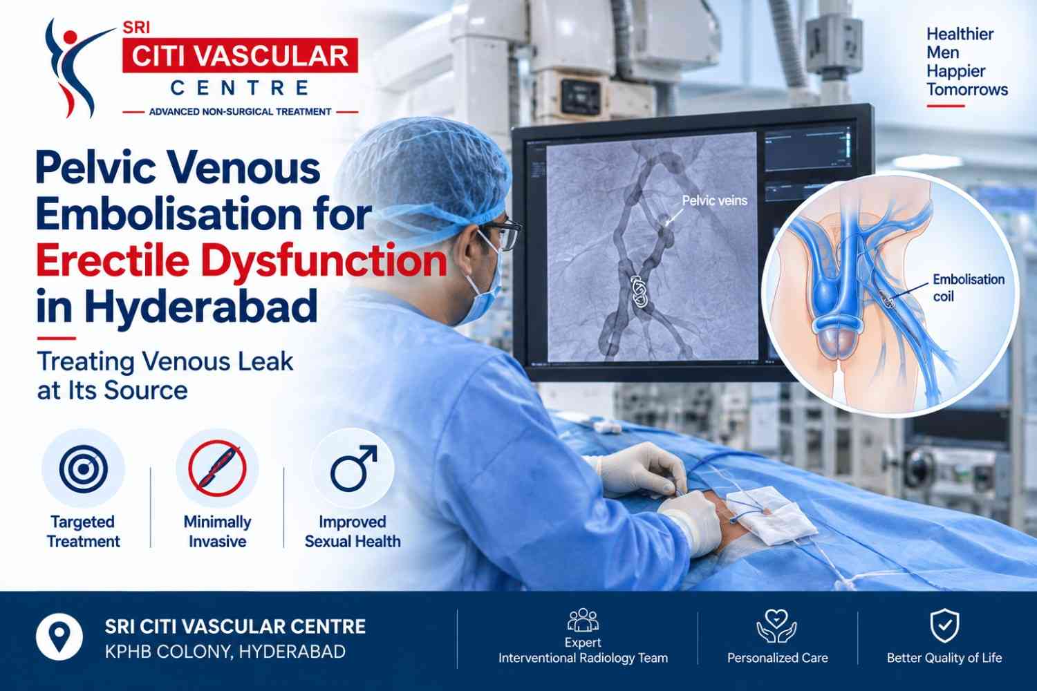
Pelvic venous embolisation is an endovascular procedure — performed entirely through small vascular access points under imaging guidance, without any open surgical incision on the penis, scrotum, or perineum. The procedure is performed by an Interventional Radiologist in a dedicated vascular imaging suite equipped with fluoroscopy and digital subtraction angiography.
The principle is conceptually straightforward: identify the specific venous channels responsible for abnormal venous outflow from the corpus cavernosum — using the CT cavernosography map obtained during assessment — and selectively close those channels with embolic material delivered through a catheter. The goal is targeted occlusion of the pathological draining veins while preserving normal and non-target venous structures. What makes the procedure technically demanding is the anatomy: the penile venous system is small-calibre, variable between individuals, and operates under different haemodynamic conditions depending on the degree of tumescence — requiring operator familiarity with this specific anatomical territory.
The procedural approach varies between centres and depends on the patient's venous anatomy, the access route selected, and the specific draining veins identified on CT cavernosography. The following describes the most commonly published approach — anterograde access via the deep dorsal penile vein — alongside the alternative pelvic venous retrograde access route.
|
1 |
Pre-Procedure Review and Consent CT cavernosography, pharmacological Doppler report, IIEF baseline score, hormonal profile, and clinical history are reviewed by Dr. Garge. The venous anatomy is mapped — identifying the dominant abnormal drainage pathways, the access route, and the embolic strategy. Consent is completed with specific discussion of: the realistic success rate (59.4% clinical improvement in the largest registry), the recurrence risk (10.2%), the pulmonary embolism risk (3/175 in the 2025 registry), and the possibility that symptoms may not improve despite technically successful embolisation. |
|
2 |
Vasoactive Injection and Access A vasoactive agent (PGE1 or tri-mix) is injected intracavernosally under sterile conditions to produce tumescence — for anterograde deep dorsal vein access, adequate tumescence is needed to fill and dilate the dorsal venous system for access. The deep dorsal penile vein — which runs along the dorsal midline of the penis, deep to Buck's fascia — is accessed through a small direct puncture under ultrasound guidance. A small-calibre sheath is introduced. For the alternative retrograde approach: transfemoral venous access is used from the groin, advancing through the femoral vein → iliac vein → internal pudendal venous system. |
|
3 |
Venography — Mapping the Drainage Pathways Under Fluoroscopy With the sheath in position, contrast is injected under digital subtraction fluoroscopy to create a detailed venogram. This maps the venous drainage from the corpus cavernosum in real time under tumescent conditions — showing: the deep dorsal vein course, cavernous veins running posteriorly, connections to the internal pudendal venous plexus, periprostatic venous channels, and any accessory aberrant draining veins. The fluoroscopic venogram is compared with the pre-procedure CT cavernosography map to confirm the anatomy and plan the selective catheterisation. |
|
4 |
Selective Catheterisation of Abnormal Drainage Channels A microcatheter is advanced selectively through the sheath into the specific abnormal venous channels identified on venography. The objective is to position the catheter tip within the target vein — not at the access point — so that embolic material is delivered precisely into the abnormal drainage pathway rather than at a non-specific proximal location. The microcatheter approach allows selection of small-calibre cavernous veins and accessory channels that standard catheters cannot reach. |
|
5 |
Embolisation — Selective Occlusion of Abnormal Veins The selected embolic agent is delivered through the microcatheter. Fluoroscopic monitoring throughout the embolisation confirms the material is staying within the target vein and is not refluxing into non-target structures. The operator monitors for any evidence of non-target embolisation — the appearance of embolic material in the periprostatic plexus, bladder veins, or, critically, the iliac veins (which communicate with the pulmonary circulation). Embolisation continues until adequate occlusion is confirmed on repeat venography. Multiple draining veins may require sequential catheterisation and embolisation. |
|
6 |
Completion Venography and Closure A completion venogram after all planned embolisation confirms adequate occlusion of targeted draining veins and the absence of significant non-target embolisation. The access sheath is removed and the access site closed. The patient is monitored for 4–6 hours after the procedure. Respiratory status is assessed as part of post-procedural observation — the PE risk, though rare, is most likely to be detected in the post-procedure monitoring period. |
Book your Appointment here:
|| Appoinment: || Call +91-7032508468 || Whatsapp:+91-7337583901 || Citi Vascular Hospital, KPHB Colony, Hyderabad.||
There is no single universally correct embolic agent for pelvic venous embolisation in erectile dysfunction. The choice depends on the target vein's calibre, flow characteristics, access route, and the operator's technique. Published series have used several different agents, often in combination.
|
Embolic Material |
Mechanism |
Used For in Penile Venous Embolisation |
|
Liquid embolic agents (Onyx / NBCA glue) |
Liquid fills the venous channel and polymerises in situ, creating a cast of the treated vessel |
Small-calibre cavernous veins and accessory drainage channels — reaches vessels that coils cannot fill adequately. Published series for venogenic ED predominantly use liquid embolic agents. |
|
Metallic coils (pushable or detachable) |
Deployed into target vein — promotes thrombosis and fibrous occlusion |
Larger pudendal vein segments. Detachable coils allow repositioning before deployment for precision placement. |
|
Vascular plugs (Amplatzer-type) |
Occlusion of a larger-calibre venous segment with a mechanical plug |
Proximal pudendal vein trunk when a larger-calibre target vessel is present. Less commonly used as primary agent for penile venous embolisation. |
|
Sclerosant agents |
Chemical agent injected into target vein — causes endothelial damage and thrombotic occlusion |
Selected venous channels in some published case series. Less commonly used as the primary technique in published endovascular ED registries. |
|
Combined approach |
Multiple agents used sequentially within the same procedure |
Most commonly required in practice — larger veins with coils or plugs, smaller channels with liquid embolic agents. |
Pelvic venous embolisation for erectile dysfunction is an evolving field with a growing but still predominantly observational evidence base. Patients and clinicians should understand both what the published data shows and the important caveats that apply to interpreting it.
|
Study / Source |
Key Findings and Clinical Implications |
|
CVIRENDO Registry. Cardiovasc Intervent Radiol. 2023. (50 patients, 6-week follow-up) |
First prospective registry specifically for venogenic ED embolisation. Technical success: 98%. Clinical improvement meeting predefined feasibility endpoint: 68% at 6 weeks. Access: anterograde deep dorsal penile vein. Embolic agent: liquid embolic (Onyx). Established proof of concept and early efficacy data for the anterograde approach. |
|
Multicentre Registry. Cardiovasc Intervent Radiol. 2025. (175 patients, ~9-month follow-up) |
Most comprehensive published registry to date. Technical success: 99.5%. Clinical improvement (predefined endpoint at follow-up): 59.4%. Recurrence or persistence of venous leak: 10.2%. Non-target embolisation (PE): 3 patients — no long-term consequences reported in those cases. Important: the 59.4% improvement rate reflects a predefined endpoint at approximately 9 months — some patients improve beyond this timeframe; individual outcomes vary. |
|
Endovascular therapy for erectile dysfunction — > 1000 treatments. CVIR Endovascular. 2026. |
Largest published endovascular ED experience including venous and arterial cases. Confirms the growing real-world experience base and supports the technical feasibility of venous embolisation at dedicated endovascular ED centres. |
|
Venogenic ED via deep dorsal penile vein access — CT cavernosography and endovascular treatment. J Med Case Reports. 2022. |
Case series describing the anterograde access technique and the role of CT cavernosography in planning — confirming that imaging-guided selection produces better technical and clinical outcomes than clinical selection alone. |
What the 59.4% improvement figure means: In the 2025 multicentre registry of 175 patients, 59.4% met the predefined clinical improvement endpoint at approximately 9 months. This does not mean 59.4% were cured or achieved natural erections without medication. It means 59.4% met the study's specific threshold for meaningful clinical improvement — reduction in IIEF scores, change in EDV on Doppler, or patient-reported improvement — at the study timepoint. Some patients in the 40.6% non-responder group had technically successful embolisation confirmed on imaging: the venous leak was closed, but erectile function did not improve — because other mechanisms (arterial disease, neurogenic, cavernous smooth muscle atrophy) were also contributing. This is why proper diagnostic assessment before patient selection matters more than any procedural technical detail.
Pelvic venous embolisation is a minimally invasive procedure — but 'minimally invasive' does not mean risk-free. Patients should have a complete and honest understanding of the risk profile before making a decision.
|
Complication |
Frequency |
Clinical Notes |
|
Access site pain or bruising |
Common — expected |
Discomfort at the dorsal penile vein access site or perineal region for 2–5 days. Managed with paracetamol and NSAIDs. Self-limiting. |
|
Post-embolisation scrotal/perineal aching |
Common — expected |
Mild aching in the scrotal or perineal region for 3–7 days as the embolised venous territory responds to occlusion. Self-limiting in most cases. |
|
Non-target embolisation — pulmonary embolism |
Rare — 3/175 (1.7%) in 2025 registry |
The most serious complication. Embolic material entering the deep dorsal vein or pudendal veins can, via venous connections, reach the pulmonary circulation. In the 2025 multicentre registry (175 patients), PE occurred in 3 patients — no long-term consequences were reported in those cases. Post-procedure respiratory monitoring is an essential safety step. Risk is reduced by careful fluoroscopic monitoring during embolisation and avoidance of non-selective bulk injection. |
|
Recurrence of venous leak |
10.2% in 2025 registry |
Persistent or recurrent venous outflow at follow-up — either from recanalisation of embolised veins or development of new collateral drainage channels. Repeat embolisation may be considered. |
|
Failure to improve erectile function |
~40% of cases |
Despite technically successful venous occlusion, erectile function may not meaningfully improve. Most commonly because co-existing mechanisms — arterial insufficiency, cavernous smooth muscle atrophy, neurogenic component — were not identified pre-procedurally or were under-weighted in the selection decision. |
|
Penile vein thrombosis |
Possible — access site |
Thrombosis at the deep dorsal penile vein access site. Usually resolves with anticoagulation if clinically significant. Prevented by procedural technique and post-procedure anticoagulation protocol as per operator's plan. |
|
Infection |
Rare — < 1% |
Procedural infection at the access site. Prevented by sterile technique. Managed with antibiotics if it occurs. |
Pulmonary embolism — patient communication: The 3/175 PE incidence (1.7%) in the 2025 multicentre registry represents a specific risk that patients must understand before consenting. The penile and pudendal venous system communicates with the internal iliac venous system, which drains into the iliac veins and inferior vena cava. Embolic material that refluxes proximally can enter this pathway and reach the pulmonary circulation. The 2025 registry authors note that no long-term consequences occurred in the 3 affected patients — suggesting subclinical or small PE in those cases. However, pulmonary embolism carries a mortality risk in some circumstances. This risk should be explicitly discussed at consent, and post-procedure respiratory monitoring performed as standard. Call +91-73375 83901 to discuss.
|
Timeframe |
What to Expect |
|
During procedure |
Awake — local anaesthesia and IV sedation. Pressure at the access site. No pain from the embolisation itself (veins are not pain-sensitive). Duration: typically 60–90 minutes depending on anatomy complexity and number of vessels treated. |
|
Immediately after (0–6 hours) |
Monitored in day-care unit — vital signs, respiratory rate, oxygen saturation (PE monitoring), and access site check. Most patients are well and discharged the same day after satisfactory observation. |
|
Day 1–5 |
Mild perineal, scrotal, or penile shaft discomfort — the most common post-procedure symptom. Managed with paracetamol and NSAIDs. Avoid sexual activity for 2 weeks initially. Avoid heavy lifting, strenuous physical activity, and prolonged seated pressure on the perineum. |
|
Week 2–6 |
Return to desk work typically within 3–7 days. Light activity resumable from Day 5. Sexual activity may be attempted from Week 2–4 as directed by the treating team. PDE5 inhibitors may be used during this assessment period — combination with embolisation may improve outcomes in some patients. |
|
Month 1–3 |
First formal assessment of erectile function improvement. IIEF score repeated. Pharmacological penile Doppler performed to confirm reduction in EDV and improved RI — objective confirmation that the veno-occlusive mechanism has been enhanced. Many patients notice improvement beginning 4–6 weeks after the procedure; peak improvement assessed at 3–6 months. |
|
Month 6–9 |
The 2025 multicentre registry uses approximately 9 months as its primary endpoint — aligning with when full treatment effect is typically assessed. A second IIEF and Doppler confirm durability. If venous leak has recurred (EDV elevated again on follow-up Doppler), repeat embolisation is discussed. |
Yes — and patients must understand this before proceeding. The 2025 multicentre registry (175 patients) reported recurrence or persistence of venous leakage in 10.2% of patients during its follow-up period. This is not unique to this procedure — all treatments for venous leak (including the largely abandoned surgical venous ligation) carry a recurrence risk as the vascular system develops collateral drainage channels around the treated vessels.
Two mechanisms drive recurrence: recanalization — the embolised vein re-opens as the embolic material is partially absorbed or the thrombus organised; and collateral development — new venous channels emerge to take over the drainage function of the embolised vessel. The embolisation technique can influence the first mechanism (liquid embolic agents generally produce more durable occlusion than coils alone), but cannot prevent collateral formation. If venous leak recurs and is confirmed on follow-up Doppler, repeat embolisation using the same or modified technique is generally technically feasible and is the appropriate next step in most cases.
|
Feature |
Pelvic Venous Embolisation |
Surgical Venous Ligation (largely abandoned) |
|
Access |
Percutaneous — small vascular access point | No open incision |
Open perineal or scrotal surgical incision | General/spinal anaesthesia |
|
Venous Anatomy Addressed |
Multiple draining channels — cavernous veins, accessory veins, deep dorsal vein — based on CT cavernosography |
Primarily the deep dorsal vein — misses cavernous and accessory draining channels which are the primary pathological vessels in most patients |
|
Recurrence |
10.2% in 2025 registry | Repeat embolisation feasible |
High recurrence as new collateral channels develop — reason why surgical ligation has been largely abandoned at most urology centres |
|
Recovery |
Same-day discharge | 3–7 days to desk work |
2–5 days hospital stay | 2–4 weeks recovery |
|
Compared to Penile Prosthesis |
Natural erection mechanism preserved | Reversible | 59.4% improvement in registry | Not guaranteed to work |
Surgical prosthesis: > 90% satisfaction | Reliable and guaranteed rigidity | Irreversible | Natural tissue permanently altered |
The correct position of pelvic venous embolisation in the ED treatment hierarchy is: after medications and non-invasive treatments have been tried and found insufficient; before penile prosthesis in younger men who prefer a natural-function approach. It is not a replacement for penile prosthesis when embolisation has failed and other treatments have been exhausted — in that scenario, prosthesis remains the most reliable final option.
Q1: What is pelvic venous embolisation for erectile dysfunction?
A: Pelvic venous embolisation is a minimally invasive endovascular procedure that selectively closes the abnormal veins responsible for excessive venous outflow from the corpus cavernosum — the vascular mechanism of venous leak erectile dysfunction. A catheter is advanced to the target venous pathways (typically via the deep dorsal penile vein or pelvic venous access) under fluoroscopic guidance, and embolic material is delivered to occlude the abnormal drainage channels. No open surgery. No penile incision. 2025 registry (175 patients): 99.5% technical success, 59.4% clinical improvement at ~9 months.
Q2: How is venous leak erectile dysfunction diagnosed before embolisation?
A: Three-step assessment: (1) Clinical history, IIEF score, hormonal profile. (2) Pharmacological penile Doppler after intracavernosal PGE1 injection — EDV > 5 cm/s + RI < 0.75 at peak tumescence with adequate PSV (> 25 cm/s) confirms venous leak. (3) CT cavernosography — contrast injected into the corpus cavernosum under tumescence maps the deep dorsal vein, cavernous veins, and pudendal vein drainage pathways, confirming which veins are anatomically treatable by embolisation. Symptoms alone cannot diagnose venous leak.
Q3: What is CT cavernosography?
A: CT cavernosography is the key anatomical investigation for planning venous embolisation for erectile dysfunction. After intracavernosal injection of a vasoactive agent (to produce maximal tumescence) and contrast material, CT images map the entire penile and pelvic venous drainage system under functional tumescent conditions. It identifies the deep dorsal vein, cavernous veins, pudendal veins, periprostatic channels, and any accessory draining veins — allowing the interventional radiologist to plan exactly which veins to target during embolisation and which access route to use.
Q4: What access route is used for pelvic venous embolisation?
A: The most commonly published access route is anterograde via the deep dorsal penile vein — a direct puncture into the deep dorsal vein on the dorsal midline of the penis under ultrasound guidance after a vasoactive injection produces tumescence. This anterograde approach follows the natural venous flow direction from the penis toward the pelvis and allows selective catheterisation of the cavernous veins and downstream pathways. An alternative retrograde approach via transfemoral pelvic venous access is also used — the access choice depends on anatomy and operator expertise.
Q5: What is the success rate of pelvic venous embolisation for erectile dysfunction?
A: The most comprehensive published evidence is the 2025 multicentre registry of 175 patients: technical success 99.5%, predefined clinical improvement endpoint met at approximately 9 months: 59.4%, recurrence/persistence of venous leak: 10.2%. These figures should not be interpreted as guaranteed outcomes for an individual patient — they represent registry averages across varying patients and centres. The 2023 single-centre registry of 50 patients reported 68% improvement at 6 weeks. These results are encouraging but improvement is not guaranteed.
Q6: Is there a risk of pulmonary embolism from venous embolisation for ED?
A:Yes — this is a specific risk that requires honest discussion before consent. In the 2025 multicentre registry (175 patients), pulmonary embolism occurred in 3 patients (1.7%) as a consequence of non-target embolisation — embolic material entering the pulmonary circulation via the penile and pudendal venous connections with the iliac venous system. The authors reported no long-term consequences in those 3 cases. Post-procedure respiratory monitoring is performed as standard at Citi Vascular Centre to detect this complication early. The risk is minimised by careful fluoroscopic monitoring during embolisation and avoidance of non-selective bulk injection.
Q7: Does everyone who cannot maintain an erection have venous leak?
A:No — and this is one of the most important clinical points on this page. Difficulty maintaining an erection occurs with arterial insufficiency, diabetes, medication side effects, Peyronie's disease, neurological conditions, post-prostatectomy changes, and psychological/performance anxiety — none of which are venous leak and none of which would benefit from venous embolisation. A proper diagnostic assessment (pharmacological Doppler + CT cavernosography) is essential to confirm that the dominant mechanism is genuinely venogenic before embolisation is considered.
Q8: Can venous leak return after embolisation?
A: Yes — the 2025 multicentre registry reported recurrence or persistence in 10.2% of patients. Recurrence occurs through two mechanisms: recanalisation (embolised vein re-opens) and collateral development (new venous channels emerge). Liquid embolic agents generally produce more durable occlusion than coils alone. If venous leak recurs and is confirmed on follow-up Doppler, repeat embolisation is technically feasible in most cases. Patients should understand this limitation before the first procedure.
Q9: Will I still need Viagra or Cialis after venous embolisation?
A: Possibly — the goal of embolisation is to improve the veno-occlusive mechanism, which may allow PDE5 inhibitors that were previously ineffective to become effective. Some patients who respond well to embolisation find they need less medication or lower doses. Others continue to use PDE5 inhibitors after embolisation, now finding them more effective than before. A minority of well-selected patients may achieve adequate erections without medication. The expectation should be improved erectile function — not automatic elimination of all medication.
Q10: How long does the procedure take and is it painful?
A: The procedure typically takes 60–120 minutes — depending on the number of abnormal venous channels requiring treatment, the complexity of the venous anatomy, and whether re-catheterisation of additional channels is needed. It is performed under local anaesthesia + IV sedation. The patient is awake and comfortable throughout. The access site is numbed with local anaesthetic. The embolisation itself is generally not painful because veins are not pain-sensitive. Mild perineal or penile shaft discomfort in the 3–7 days after the procedure is common and is managed with paracetamol.
Q11: When should I see results after pelvic venous embolisation?
A: Improvement in erectile function is gradual — not immediate. Most patients begin noticing meaningful improvement 4–8 weeks after the procedure. The 2025 multicentre registry uses approximately 9 months as its primary endpoint — consistent with the timeframe for full embolisation effect and venous fibrous occlusion. A follow-up pharmacological Doppler at 3 months confirms objective improvement (reduced EDV, improved RI). A PDE5 inhibitor trial from 4–6 weeks after the procedure may reveal improved response in men who were previously non-responders.
Q12: Who performs pelvic venous embolisation for erectile dysfunction in Hyderabad?
A: Dr. Shaileshkumar Garge — FRCR (UK), FNVIR (CMC Vellore), EBIR (Spain) — Director and Chief Vascular Physician at Citi Vascular Centre, KPHB Colony, Hyderabad, performs pelvic venous embolisation for venous leak erectile dysfunction. The complete diagnostic pathway — pharmacological penile Doppler, CT cavernosography, and embolisation — is available under one specialist. All consultations are strictly confidential. Call +91-73375 83901 or WhatsApp 73375 83901.
|
Reference |
Key Finding |
|
CVIRENDO Registry. Cardiovasc Intervent Radiol. 2023. (50 patients) |
Prospective registry — anterograde deep dorsal penile vein access with liquid embolic (Onyx). Technical success 98%. Clinical improvement 68% at 6 weeks using predefined feasibility endpoint. Established proof of concept for the anterograde approach. |
|
Multicentre Registry. Cardiovasc Intervent Radiol. 2025. (175 patients) |
Most comprehensive published registry. Technical success 99.5%. Clinical improvement 59.4% at approximately 9 months. Recurrence/persistence 10.2%. PE: 3/175 (1.7%) — no long-term consequences. Key safety and efficacy reference for patient counselling. |
|
Endovascular therapy for erectile dysfunction — > 1,000 treatments. CVIR Endovascular. 2026. |
Largest published endovascular ED experience. Confirms real-world feasibility and growing experience base for venous and arterial endovascular approaches to erectile dysfunction. |
|
Venogenic ED — CT cavernosography and deep dorsal vein access. J Med Case Reports. 2022. |
Case series confirming that CT cavernosography provides the anatomical information required for appropriate patient selection and technical planning — differentiating patients with treatable venous anatomy from those where embolisation is unlikely to be complete. |
|
EAU Guidelines on Sexual and Reproductive Health — Erectile Dysfunction. 2024. |
Acknowledges venous embolisation as an emerging endovascular option for veno-occlusive dysfunction in selected patients, within the evidence-based ED treatment algorithm. |
Citi Vascular Centre, KPHB Colony, Road No. 1, Hyderabad — pelvic venous embolisation for venous leak ED for patients from:
Kukatpally and KPHB — 5 min | Miyapur and Bachupally — 10 min
Hitech City, Ameerpet and Madhapur — 20 min | Gachibowli and Banjara Hills — 25 min
Secunderabad and Begumpet — 25 min | Telangana & AP — outstation welcome
Pelvic venous embolisation for erectile dysfunction is an endovascular treatment for a specific and well-defined clinical problem: confirmed venogenic erectile dysfunction where pharmacological penile Doppler demonstrates elevated end-diastolic velocity and reduced resistive index, and CT cavernosography maps anatomically treatable abnormal venous drainage pathways. It is not a treatment for every man who cannot maintain an erection. The symptom pattern of venous leak — erection that initiates but rapidly deflates — is shared by multiple other causes of erectile dysfunction that would not benefit from embolisation.
When properly selected patients undergo the procedure, the published evidence is encouraging: the 2025 multicentre registry (175 patients) reports 99.5% technical success and 59.4% meeting a predefined clinical improvement endpoint at approximately 9 months. The 10.2% recurrence rate and the 1.7% pulmonary embolism incidence are important known risks that must be disclosed at consent. The procedure is minimally invasive — same-day discharge, no open surgery, no penile incision — and can be repeated if recurrence occurs. For younger men with confirmed venous leak who have failed adequate PDE5 inhibitor trials, pelvic venous embolisation offers a catheter-based approach to improving erectile function before committing to the irreversible option of penile prosthesis surgery.
At Citi Vascular Centre, KPHB Colony, Hyderabad, Dr. Garge provides the complete venous ED diagnostic and treatment pathway — pharmacological Doppler, CT cavernosography, and selective pelvic venous embolisation using liquid embolic agents, coils, and combined technique as appropriate to the patient's venous anatomy. All consultations are strictly confidential.
Book your Appointment here:
|| Appoinment: || Call +91-7032508468 || Whatsapp:+91-7337583901 || Citi Vascular Hospital, KPHB Colony, Hyderabad.||