Bronchial Artery Embolisation in Hyderabad (2026) | BAE for Haemoptysis
|
TABLE OF CONTENTS |
|
1. |
Introduction + Quick Answer |
7. |
Success Rates and Recurrence |
|
2. |
Quick Facts |
8. |
Risks and Complications |
|
3. |
What Is Haemoptysis and When Is It an Emergency? |
9. |
BAE vs Surgery |
|
4. |
Common Causes of Haemoptysis Requiring BAE |
10. |
Recovery After BAE |
|
5. |
What Is Bronchial Artery Embolisation? |
11. |
FAQ — 10 Q&As |
|
6. |
Step-by-Step BAE Procedure — 7 Steps |
12. |
Evidence References + Key Points + Summary |
|
1. INTRODUCTION + QUICK ANSWER |
|
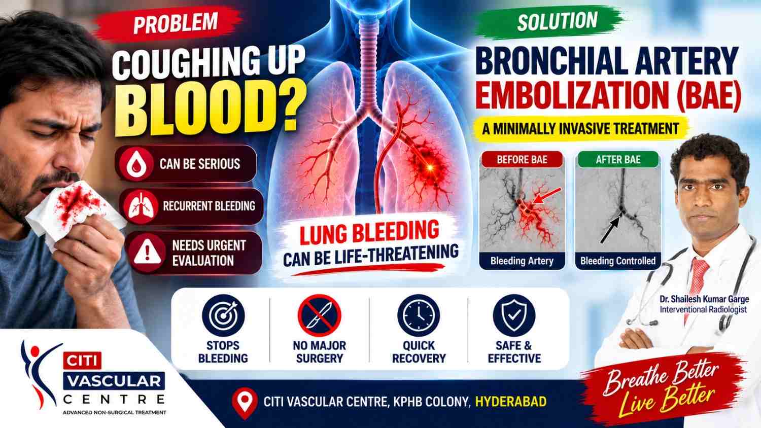
QUICK ANSWER What Is Bronchial Artery Embolisation and Who Needs It? Bronchial artery embolisation (BAE) is a minimally invasive, catheter-based interventional radiology procedure used to control haemoptysis — coughing up blood — by blocking the abnormal bronchial arteries supplying the bleeding source in the lung. It is performed under fluoroscopic guidance, requires no open surgery, and is the first-line interventional treatment for massive or recurrent haemoptysis when medical management alone is inadequate. Dr. Garge FRCR (UK) | Citi Vascular Centre, KPHB, Hyderabad | +91-73375 83901. |
Coughing up blood — haemoptysis — is always alarming. In small amounts, it may indicate an irritated airway or minor infection. In large quantities, it is a medical emergency that can fill the airways faster than a patient can breathe, causing asphyxia rather than simple blood loss. For the patients who experience massive or recurrent haemoptysis — often those with tuberculosis, bronchiectasis, aspergilloma, or lung cancer — the traditional options were limited: aggressive medical stabilisation while hoping the bleeding would stop, or high-risk emergency thoracic surgery on a compromised patient.
Bronchial artery embolisation changed this equation significantly. By identifying and blocking the specific bronchial artery feeding the bleeding source through a minimally invasive catheter approach, interventional radiology can stop haemoptysis rapidly in most patients without the risks of open-chest surgery. BAE is now the first-line interventional treatment for massive and recurrent haemoptysis at specialist vascular centres worldwide. This guide explains how it works, who needs it, the step-by-step procedure, what to expect during recovery, and the honest success rates and limitations.
|
Bronchial Artery Embolisation — Citi Vascular Centre, KPHB, Hyderabad Call +91-73375 83901 | WhatsApp 73375 83901 | citivascularcentre.com | Mon–Sat 9AM–6PM |
|
2. QUICK FACTS — BRONCHIAL ARTERY EMBOLISATION |
|
Feature |
Detail |
|
Procedure Type |
Minimally invasive catheter-based embolisation — no open surgery, no thoracotomy |
|
Guidance |
Fluoroscopy + digital subtraction angiography (DSA) throughout |
|
What It Treats |
Massive haemoptysis (> 200–300mL/24hr) | Recurrent haemoptysis | Haemoptysis uncontrolled by medical treatment |
|
Access Route |
Transfemoral (groin) — arterial approach via femoral artery → aorta → bronchial artery |
|
Anaesthesia |
Local anaesthesia at groin access site + IV sedation | General anaesthesia rarely required |
|
Immediate Control |
Haemoptysis arrested in 73–98% of cases at initial session — published systematic review data |
|
Recurrence |
Varies by underlying cause: 10–55% within months to years. Underlying disease management is critical. |
|
Hospital Stay |
2–5 days typically — depending on the severity of haemoptysis, underlying diagnosis, and post-procedure stability |
|
Most Critical Risk |
Spinal cord ischaemia from non-target embolisation of spinal arteries — rare (< 1%) but serious. Requires meticulous angiographic technique. |
|
3. WHAT IS HAEMOPTYSIS AND WHEN IS IT AN EMERGENCY? |
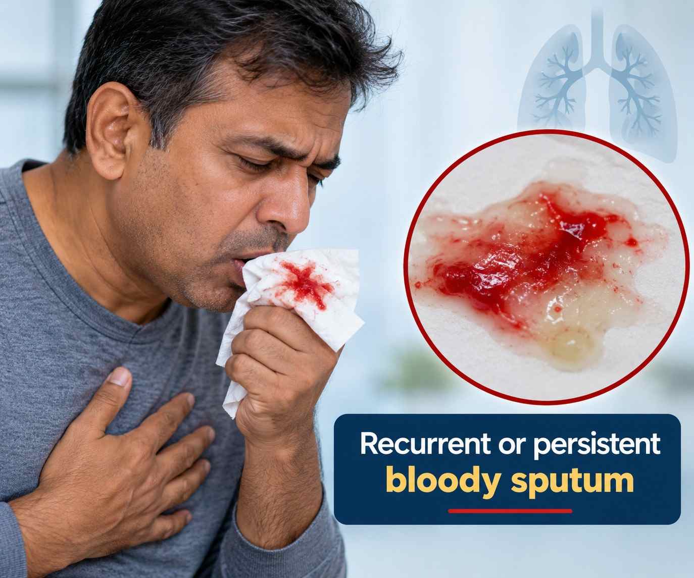
Haemoptysis is the coughing up of blood originating from the respiratory tract — specifically from the lungs and the airways below the larynx. Distinguishing true haemoptysis (lower respiratory origin) from haematemesis (vomiting blood from the upper gastrointestinal tract) and pseudo-haemoptysis (blood from the nasopharynx draining into the airway) is clinically important but can be challenging acutely.
|
Volume |
Clinical Classification and Urgency |
|
Blood-streaked sputum |
Common — usually minor bronchial vessel irritation, mucosal infection, or minor bronchiectasis. Warrants investigation but is not immediately life-threatening. |
|
Moderate haemoptysis (30–200mL/day) |
Requires hospital assessment, investigation for underlying cause, and monitoring. Haematological stability typically preserved. Bronchoscopy and CT to identify source. |
|
Massive haemoptysis (> 200–300mL/day or any haemoptysis causing airway compromise) |
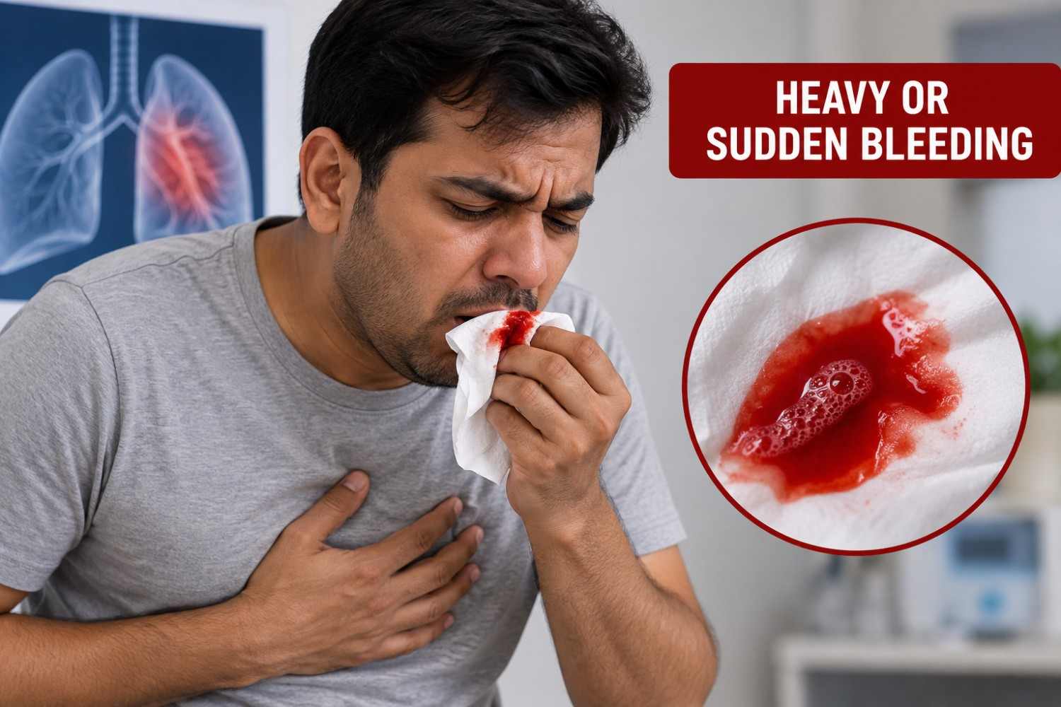
Medical emergency. Risk of asphyxia — death in massive haemoptysis is usually from drowning in one's own blood, not blood loss. Immediate haemodynamic stabilisation, airway protection, and urgent BAE referral. |
|
When to seek emergency care immediately: Large volumes of bright red blood coughed up (more than a small cup) | Haemoptysis with breathlessness or inability to clear airways | Haemoptysis with sudden drop in blood pressure | Known lung disease (TB, bronchiectasis, aspergilloma) with new or worsening haemoptysis | Any haemoptysis after a procedure or trauma. Call emergency services and inform the team. Do not lie flat — sitting upright helps keep the airway clear. |
|
4. COMMON CAUSES OF HAEMOPTYSIS REQUIRING BAE |
The distribution of haemoptysis causes varies significantly by geography. In India and Southeast Asia, tuberculosis (TB) — both active and post-TB sequelae — accounts for a very large proportion of significant haemoptysis. Understanding the underlying cause is important not only for BAE planning but because BAE controls bleeding but does not treat the underlying disease.
|
Cause |
Clinical Notes |
|
Tuberculosis (TB) — active and post-TB |
Most common cause of massive haemoptysis in India. Rasmussen's aneurysm (pseudoaneurysm in TB cavities) and bronchiectasis from prior TB are frequent sources. Bronchial arteries become massively hypertrophied and tortuous. |
|
Bronchiectasis |
Chronic airway dilation — bronchial arteries hypertrophy as part of chronic inflammatory response. Major source of recurrent haemoptysis across all age groups. |
|
Aspergilloma (fungal ball) |
Fungal colony in a pre-existing cavity — typically post-TB. The surrounding tissue is highly vascularised. Associated with particularly significant haemoptysis episodes. |
|
Lung cancer |
Both primary bronchogenic carcinoma and metastatic disease can cause haemoptysis through direct vessel invasion or erosion. BAE can palliate bleeding in non-surgical candidates. |
|
Chronic lung abscess |
Cavitating infection with necrosis of pulmonary parenchyma — surrounding inflammatory bronchial artery hypertrophy produces bleeding. |
|
Arteriovenous malformation (AVM) |
Pulmonary AVM — direct shunt between pulmonary artery and vein — can cause haemoptysis. Distinct from BAE indication: usually managed by pulmonary artery side embolisation, not bronchial artery. |
|
5. WHAT IS BRONCHIAL ARTERY EMBOLISATION (BAE)? |
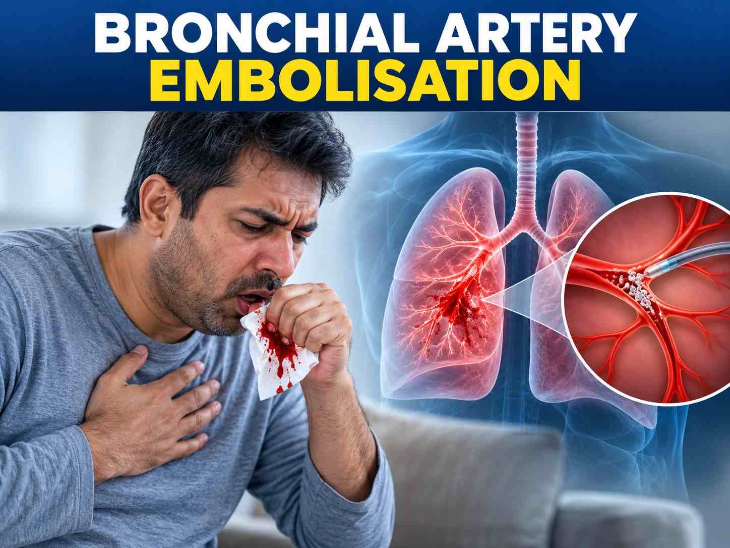
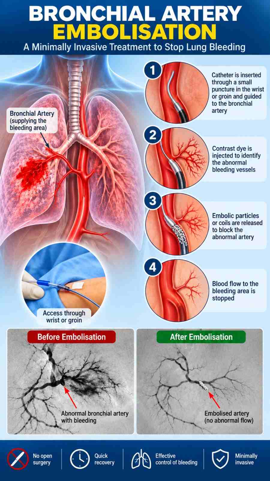
Bronchial artery embolisation is a catheter-based procedure in which the interventional radiologist selectively catheterises the bronchial arteries feeding the bleeding source in the lung and injects embolic material to block them — stopping the abnormal blood flow that is causing the haemoptysis. The bronchial arteries are the systemic-pressure supply to the airway and lung parenchyma. When they become hypertrophied, tortuous, and abnormally vascular as part of chronic disease, they are the source of the vast majority of significant haemoptysis episodes.
The critical technical principle underlying BAE is selective embolisation — blocking only the specific bronchial arteries responsible for the bleeding, while preserving flow in non-target vessels, particularly any spinal artery branches that share origin with the bronchial artery. The artery of Adamkiewicz and anterior spinal artery branches arising from intercostal or bronchial origins must be identified and specifically avoided during embolisation — non-target embolisation of these vessels is the mechanism of spinal cord ischaemia, the most serious possible complication of BAE.
|
6. STEP-BY-STEP BAE PROCEDURE — 7 STEPS |
The following describes the BAE procedure as performed at Citi Vascular Centre, KPHB, Hyderabad. The procedure requires the full capability of a dedicated vascular interventional suite with high-quality digital subtraction angiography, a well-stocked embolic material inventory, and an experienced operator with anatomical knowledge of bronchial artery variants.

|
1 |
Pre-Procedure Preparation CT pulmonary angiography (CTPA) or CT chest is reviewed before the procedure to identify the side and approximate location of bleeding, any cavities, bronchiectatic changes, or masses. Chest X-ray, recent bronchoscopy reports, and haematological status (coagulation, platelet count, haemoglobin) are assessed. IV access established, oxygen applied, and the patient is stabilised haemodynamically as much as possible. |
|
2 |
Arterial Access A small puncture is made in the femoral artery in the groin under local anaesthesia and sterile conditions. A vascular sheath is inserted. This provides the access route for all subsequent catheter work. Haemostasis at the end of the procedure is achieved by manual pressure, suture-based closure, or a vascular closure device. |
|
3 |
Aortography and Bronchial Artery Identification A flush catheter is positioned in the descending thoracic aorta and a digital subtraction aortogram is performed. This provides a road map of the bronchial artery origins — their positions relative to the vertebral bodies, the number of bronchial arteries, and whether any common trunks with intercostal arteries are present (intercostobronchial trunks). Bronchial arteries typically arise from the descending thoracic aorta at T5–T6 level but significant anatomical variation is common. |
|
4 |
Selective Bronchial Artery Catheterisation A shaped catheter (or microcatheter for smaller vessels) is advanced into each target bronchial artery. The position is confirmed on fluoroscopy and contrast injection. The arterial anatomy is carefully assessed — particularly for spinal artery branches. Any vessel showing a spinal artery origin (hairpin turn pattern, or vessels feeding the spinal canal) must not be embolised. |
|
5 |
Diagnostic Angiography Selective contrast injection maps the bronchial artery territory — showing hypertrophy, tortuosity, and abnormal vascularity consistent with the bleeding source. Features suggesting the target vessel include: markedly enlarged calibre | tortuous 'corkscrew' appearance | hypervascularity in the affected lung region | active contrast extravasation (if actively bleeding). Comparison with the unaffected side helps identify abnormal vessels. |
|
6 |
Embolisation — Blocking the Target Vessels Embolic material is injected carefully through the catheter into the target bronchial artery. Particulate embolic agents (polyvinyl alcohol particles or microspheres — typically 300–500 microns) are most commonly used. They lodge in the distal vessels, cutting off blood supply to the abnormal vascular bed. Embolisation is performed under careful fluoroscopic monitoring — injection is stopped if any retrograde flow toward aorta or spinal territory is seen. Adjacent bronchial arteries and hypertrophied intercostobronchial trunks are embolised as appropriate. |
|
7 |
Post-Embolisation Assessment and Closure A final angiogram after embolisation confirms satisfactory occlusion of target vessels. The catheter and sheath are removed and haemostasis achieved at the femoral access site. The patient is moved to monitored recovery. Vital signs, respiratory status, and haemoptysis volume are closely monitored for 24–48 hours. Most patients experience significant reduction or complete cessation of haemoptysis within the first few hours after successful BAE. |
|
7. SUCCESS RATES AND RECURRENCE |
Published evidence for BAE is substantial — this procedure has been performed at specialist centres for over four decades, and systematic reviews provide a clear picture of what patients can realistically expect.
|
Outcome |
Published Evidence |
|
Immediate haemoptysis control |
73–98% across published series (systematic review: Sopko and Kim). High rates of immediate bleeding control — this is BAE's primary strength. |
|
Short-term recurrence (within 30 days) |
10–30% — more common when the underlying disease has not been treated or multiple bronchial arteries are involved. Incomplete embolisation (not treating all contributing vessels) is a technical contributor. |
|
Long-term recurrence (1–5 years) |
Up to 55% in some series — particularly in patients with aspergilloma, cystic fibrosis, and non-TB causes. TB-related haemoptysis with effective anti-TB treatment has more favourable long-term outcomes. |
|
Repeat BAE for recurrence |
Safe and effective — most patients who experience recurrence can undergo a second BAE session. Recurrence may also prompt reassessment of the underlying diagnosis and disease management. |
|
Role of underlying disease treatment |
Critical — BAE controls the bleeding but the underlying cause remains. Anti-TB treatment, antifungal therapy for aspergilloma, antibiotics for exacerbated bronchiectasis — all reduce the risk of re-bleeding after BAE. |
|
Important: BAE controls haemoptysis — it does not cure the underlying lung disease. Patients with TB must complete a full course of anti-TB treatment. Aspergilloma patients may require ongoing antifungal management or surgical resection after BAE stabilises the acute bleed. The interventional radiologist and the referring pulmonologist should work as a team around every BAE patient. Call +91-73375 83901. |
|
8. RISKS AND COMPLICATIONS |
|
Complication |
Frequency |
Clinical Notes |
|
Post-embolisation syndrome |
Common — most patients |
Fever, chest pain, and malaise for 3–5 days after BAE — expected inflammatory response to embolised tissue. Self-limiting. Managed with paracetamol and anti-inflammatories. |
|
Chest pain / pleuritic pain |
Common |
Ischaemic pain from embolised bronchial artery territory — most cases resolve within 3–7 days. |
|
Dysphagia (difficulty swallowing) |
Uncommon |
Oesophageal artery may share an origin with bronchial artery — careful technique avoids this. Usually temporary. |
|
Spinal cord ischaemia |
Rare — < 1% but serious |
The most feared complication. Caused by non-target embolisation of a spinal artery arising from the same trunk as the bronchial artery. Meticulous pre-embolisation angiography and avoiding embolisation when spinal artery origins are present are the primary preventive measures. Experienced operators and modern embolic materials reduce this risk. |
|
Vascular access complications |
Uncommon |
Femoral artery haematoma, pseudoaneurysm, or arteriovenous fistula at the groin access site — standard vascular access complications. |
|
Bronchial necrosis / bronchoesophageal fistula |
Rare |
Over-embolisation of bronchial arteries can cause ischaemia of the airway wall — avoided by selective distal embolisation and appropriate particle size selection. |
|
Failure to control haemoptysis |
2–27% of cases |
Usually due to multiple contributing vessels not all identified and embolised, or non-bronchial systemic collaterals (internal mammary, subclavian) supplying the bleeding — missed on initial angiography. |
|
9. BAE vs SURGERY FOR HAEMOPTYSIS |
|
Feature |
Bronchial Artery Embolisation |
Surgical Resection (Lobectomy / Pneumonectomy) |
|
Approach |
Minimally invasive — catheter through groin |
Open thoracotomy or VATS |
|
Anaesthesia |
Local + IV sedation |
General anaesthesia — always |
|
Immediate haemostasis |
73–98% immediate control |
Definitive when complete resection achievable |
|
Mortality risk in emergency |
Low — 0–5% |
High in emergency setting — 10–40% for emergency pneumonectomy with active bleeding |
|
Lung preservation |
Full — no lung tissue removed |
Lung tissue removed — affects long-term respiratory reserve |
|
Long-term recurrence |
Possible — 10–55% depending on cause |
Lower if complete resection of diseased segment achieved |
|
Role |
First-line — stabilises the patient, controls bleeding, allows definitive treatment planning |
Definitive — when BAE fails, recurs, or disease is localised and resectable |
The modern approach to massive haemoptysis management is stepwise: medical stabilisation → BAE as first-line interventional control → reassessment of underlying cause → definitive treatment (surgery, antifungals, anti-TB therapy) as appropriate. Emergency surgery is reserved for patients where BAE fails or is not technically feasible, and where the patient is stable enough for thoracotomy — which in the context of massive haemoptysis is often a very high-risk group.
|
10. RECOVERY AFTER BAE |
|
Timeframe |
What to Expect |
|
Hours 0–24 (post-procedure) |
Close monitoring in the ward — vital signs, respiratory status, and haemoptysis volume. Post-embolisation fever (low-grade) and chest discomfort begin. Most patients note reduction or cessation of haemoptysis within hours of successful BAE. |
|
Days 1–3 |
Post-embolisation syndrome peak — fever (typically 37.5–38.5°C), pleuritic chest pain, malaise. Managed with paracetamol and NSAIDs. Respiratory physiotherapy if patient is stable enough. Transition from IV to oral nutrition when haemoptysis has settled. |
|
Days 3–5 |
Post-embolisation syndrome settles. Most patients' haemoptysis remains controlled. Discharge planning — ensuring adequate underlying disease treatment is in place (anti-TB therapy, antifungals as appropriate). |
|
Week 2–4 |
Groin access site fully healed. Gradual return to normal activity guided by respiratory status. Follow-up imaging (CT or CXR) to assess the embolised territory and underlying disease. |
|
Long-term |
Follow-up with pulmonologist for ongoing management of TB, bronchiectasis, or aspergilloma. Return immediately to emergency care if haemoptysis recurs — repeat BAE is feasible. |
|
11. FREQUENTLY ASKED QUESTIONS |
|
Q1: What is bronchial artery embolisation? |
|
Bronchial artery embolisation (BAE) is a minimally invasive catheter-based procedure that controls haemoptysis (coughing up blood) by blocking the specific bronchial arteries supplying the bleeding source in the lung. A thin catheter is advanced from the femoral artery in the groin through the aorta to the target bronchial artery under fluoroscopic guidance, and embolic material is injected to stop the abnormal blood flow. No open surgery. No thoracotomy. Published evidence reports 73–98% immediate haemoptysis control. |
|
Q2: When is BAE used for haemoptysis? |
|
BAE is the first-line interventional treatment for: massive haemoptysis (> 200–300mL in 24 hours) threatening airway or haemodynamic stability; recurrent moderate haemoptysis uncontrolled by medical management; haemoptysis from TB, bronchiectasis, aspergilloma, lung cancer, or chronic lung abscess that requires intervention. Any haemoptysis causing breathlessness or loss of airway control requires emergency assessment. |
|
Q3: What is the most common cause of haemoptysis in India? |
|
Tuberculosis — both active TB and post-TB sequelae (cavities, bronchiectasis, aspergilloma in old cavities) — accounts for the largest proportion of significant haemoptysis in India. TB causes severe hypertrophy and tortuosity of bronchial arteries in the affected lung segments, making these vessels the dominant source of bleeding. Complete and supervised anti-TB treatment after BAE is essential to prevent haemoptysis recurrence. |
|
Q4: How is BAE performed? |
|
BAE is performed under local anaesthesia and IV sedation. A catheter is placed through the femoral artery and guided to the bronchial arteries under fluoroscopic (X-ray) guidance. Contrast injection maps the abnormal vessels. After confirming no spinal artery branches arise from the target vessel, embolic particles are injected to block blood flow. The procedure takes 1–3 hours and most patients are admitted for 2–5 days post-procedure. |
|
Q5: What is the risk of spinal cord injury from BAE? |
|
Spinal cord ischaemia is the most serious potential complication of BAE — reported in less than 1% of cases at experienced centres. It occurs when a spinal artery branch arising from the same trunk as the target bronchial artery is inadvertently embolised. Prevention requires meticulous pre-embolisation angiography to identify spinal artery origins and strict avoidance of embolising any vessel with a spinal component. This is why BAE must be performed by an experienced interventional radiologist with specific vascular anatomy expertise. |
|
Q6: Can haemoptysis return after BAE? |
|
Yes — recurrence is possible. Short-term recurrence (within 30 days) occurs in 10–30% of cases — often due to incomplete initial embolisation (not all contributing bronchial arteries identified) or recanalization of embolised vessels. Long-term recurrence (over years) occurs in up to 55% of some patient populations — particularly aspergilloma patients. The primary driver is the underlying disease: treating TB, aspergilloma, and bronchiectasis actively after BAE significantly reduces recurrence risk. Repeat BAE is feasible and effective. |
|
Q7: Is BAE better than surgery for haemoptysis? |
|
For most patients presenting with massive haemoptysis, BAE is the preferred first-line intervention over emergency surgery — because emergency thoracic surgery for massive haemoptysis carries very high mortality (10–40% for emergency pneumonectomy) while BAE achieves 73–98% immediate haemostasis with substantially lower procedural risk. Surgery may be appropriate when BAE fails, recurs despite repeat embolisation, or when the underlying disease is localised and surgically resectable with good functional reserve. |
|
Q8: What happens after BAE — post-embolisation syndrome? |
|
Post-embolisation syndrome — low-grade fever, chest pain, malaise for 3–5 days — is an expected inflammatory response to the embolised bronchial artery territory. It is not an infection and does not indicate a complication. It is managed with paracetamol and anti-inflammatory medication. Patients are monitored in hospital during this phase. Most patients feel substantially better by Day 4–5 and can be considered for discharge when haemoptysis remains controlled and post-embolisation symptoms are resolving. |
|
Q9: How long does BAE take and what is the recovery time? |
|
The BAE procedure itself takes 1–3 hours depending on the bronchial artery anatomy complexity and the number of vessels requiring embolisation. Hospital admission is typically 2–5 days. Most patients can return to light activities within 1–2 weeks. Full recovery depends on the underlying disease — patients with active TB or severe bronchiectasis will have a more prolonged respiratory rehabilitation period than the BAE procedure itself requires. |
|
Q10: Who performs BAE in Hyderabad? |
|
At Citi Vascular Centre, KPHB Colony, Road No. 1, Hyderabad, BAE is performed by Dr. Shaileshkumar Garge — FRCR (UK), FNVIR (CMC Vellore), EBIR (Spain) — Director and Chief Vascular Physician. With 12+ years of dedicated interventional radiology and 15,000+ image-guided procedures, Dr. Garge provides expert bronchial artery embolisation with meticulous angiographic technique and multi-vessel assessment to minimise the risk of spinal complications. Call +91-73375 83901 or WhatsApp 73375 83901. |
|
EVIDENCE-BASED REFERENCES |
|
Reference |
Key Finding |
|
Sopko DR, Smith TP. Bronchial artery embolization. Semin Intervent Radiol. 2011;28(1):48–62. |
Comprehensive systematic review — immediate haemoptysis control in 73–98% of cases. Establishes BAE as the established first-line interventional treatment for massive haemoptysis. |
|
Panda A, Bhalla AS, Goyal A. Bronchial artery embolization in hemoptysis — a systematic review. Diagn Interv Radiol. 2017;23(4):307–317. |
Systematic review of 22 studies — confirms high initial technical and clinical success rates. Recurrence rates vary with underlying cause. TB-related haemoptysis has better outcomes with concurrent anti-TB treatment. |
|
Kucukay F et al. Bronchial artery embolisation in patients with massive haemoptysis. Diagn Interv Radiol. 2014. |
Documents the importance of multi-vessel approach — embolisation of all contributing bronchial arteries reduces short-term recurrence. |
|
CIRSE Standards of Practice for Bronchial Artery Embolisation. |
Defines minimum technical standards, embolic material selection, spinal artery precautions, and post-procedure monitoring for BAE at CIRSE-aligned centres. |
|
KEY POINTS |
|
Haemoptysis > 200–300mL/day or any haemoptysis causing airway compromise is an emergency requiring immediate medical attention and urgent BAE referral |
|
|
TB, bronchiectasis, aspergilloma, and lung cancer are the most common causes of significant haemoptysis requiring BAE in India |
|
|
BAE achieves immediate haemoptysis control in 73–98% of cases — it is the first-line interventional treatment for massive and recurrent haemoptysis |
|
|
Spinal cord ischaemia (< 1%) is the most serious potential complication — prevented by meticulous pre-embolisation angiography and avoiding spinal artery origins |
|
|
BAE controls bleeding but does not cure the underlying disease. Anti-TB therapy, antifungals for aspergilloma, and pulmonary rehabilitation are essential after BAE |
|
|
Dr. Shaileshkumar Garge FRCR (UK) | Citi Vascular Centre, KPHB | +91-73375 83901 | WhatsApp 73375 83901 | Mon–Sat 9AM–6PM |
|
LOCATION — BAE IN HYDERABAD |
Citi Vascular Centre, KPHB Colony, Road No. 1, Hyderabad — bronchial artery embolisation for haemoptysis patients from:
Kukatpally and KPHB — 5 min | Miyapur and Bachupally — 10 min
Hitech City, Ameerpet and Madhapur — 20 min | Gachibowli and Banjara Hills — 25 min
Secunderabad and Begumpet — 25 min | Telangana, AP — outstation welcome
|
Centre |
Contact |
Appointments |
|
Citi Vascular Centre |
+91-73375 83901 |
KPHB Colony, Road No. 1, Hyderabad 500072 | Mon–Sat 9AM–6PM |
|
Emergency |
WhatsApp 73375 83901 |
For urgent or emergency haemoptysis referrals — contact immediately | Insurance assisted | Outstation welcome |
|
SUMMARY |
Bronchial artery embolisation is the cornerstone of interventional management for massive and recurrent haemoptysis — a life-threatening emergency in which blood floods the airways faster than the body can compensate. By precisely identifying and occluding the bronchial arteries responsible for the abnormal blood supply, BAE achieves immediate haemoptysis control in 73–98% of cases without the prohibitive risks of emergency thoracic surgery. In a country where TB, bronchiectasis, and aspergilloma are common, the need for a safe, effective, minimally invasive option for managing haemoptysis is acute.
BAE is not a cure — it is a critical bridge that stabilises the patient, controls life-threatening bleeding, and creates the space for definitive underlying disease treatment to begin or continue. TB patients must complete anti-TB therapy. Aspergilloma patients need antifungal treatment or eventual surgical resection. Bronchiectasis patients need pulmonary rehabilitation. The interventional radiologist and the pulmonologist work in partnership around every BAE patient — not as competing providers but as complementary members of a team. At Citi Vascular Centre, KPHB Colony, Hyderabad, Dr. Garge provides expert bronchial artery embolisation with the technical rigour this procedure demands. Call +91-73375 83901 or WhatsApp 73375 83901.
|
Bronchial Artery Embolisation for Haemoptysis — Citi Vascular Centre, KPHB, Hyderabad Fluoroscopy-Guided | Minimally Invasive | 73–98% Immediate Control | TB | Bronchiectasis | Aspergilloma Dr. Shaileshkumar Garge | FRCR (UK) | FNVIR (CMC Vellore) | EBIR (Spain) | 12+ Years | 15,000+ Procedures Call +91-73375 83901 | WhatsApp 73375 83901 | citivascularcentre.com KPHB Colony, Hyderabad | Mon–Sat 9AM–6PM | Emergency Referrals Welcome |