Needle aspiration is a minimally invasive, image-guided procedure used to remove fluid, pus, or other material from a localized collection and, in some cases, obtain a sample for laboratory analysis.
At Citi Vascular Centre, Hyderabad, image-guided needle aspiration is performed by an experienced interventional radiology team, with the technique selected according to the location, size, nature, and clinical characteristics of the collection or lesion.

|
Dr. Shaileshkumar Garge — FRCR (UK), FNVIR (CMC Vellore), EBIR (Spain) — personally reviews your imaging and plans the safest needle access route for every aspiration |
|
Both high-resolution ultrasound and CT guidance available — the right modality selected based on collection location and depth |
|
Diagnostic fluid processing as standard — every aspirated sample sent for culture, sensitivity, and relevant laboratory analysis |
|
Emergency and same-day aspiration accepted for urgent clinical situations |
|
12+ years dedicated interventional radiology | 15,000+ image-guided procedures | USFDA-approved needles and equipment |
|
Written results summary and follow-up plan at discharge | Insurance pre-auth at no extra administrative charge |
Needle aspiration is one of the simplest and most versatile procedures in interventional radiology. A thin needle is guided under real-time ultrasound or CT imaging directly into a fluid-filled collection, cyst, abscess, or tissue lesion. The fluid or cells inside are then withdrawn through the needle using a syringe — removing the material in a single session. No surgical incision is made, no tube is left inside the body, and the procedure is typically complete within 15–30 minutes under local anaesthesia.
Needle aspiration serves two important clinical purposes — and can serve both in the same session. As a diagnostic tool, it provides a fluid or cell sample for laboratory analysis — identifying bacterial organisms, cancer cells, or other pathological findings that cannot be determined from imaging alone. As a therapeutic tool, it decompresses and drains a fluid collection or abscess — relieving pressure, pain, and the source of infection.
Common examples include:
Ascitic Fluid Aspiration — Paracentesis
Paracentesis removes excess fluid from the abdominal cavity. It may be performed to investigate the cause of ascites or to relieve symptoms caused by significant fluid accumulation.
Pleural Fluid Aspiration — Thoracentesis
Thoracentesis removes fluid from the pleural space around the lungs. It may help determine why the fluid has accumulated and can relieve pressure-related symptoms such as breathlessness. Ultrasound is commonly used to guide the procedure.
Abscess Aspiration
An abscess contains infected material or pus. In selected smaller or accessible collections, needle aspiration may be considered. Larger, thick or persistent collections may be better suited to catheter drainage or another treatment.
Fluid Collection / Cyst Aspiration
Image-guided aspiration can also be considered for selected postoperative, traumatic, cystic or other localized fluid collections. The choice between aspiration and catheter drainage depends on the collection and the patient's clinical circumstances. The Society of Interventional Radiology provides standards for image-guided aspiration and drainage of abscesses and fluid collections.
|
No incision — tiny needle entry, no stitches, no surgical wound |
|
No tube left inside — procedure complete in a single session; no drain management required |
|
Local anaesthesia only — no general anaesthesia or spinal anaesthesia required |
|
Same-day discharge — patient returns home on the same day in most cases |
|
Diagnostic and therapeutic in one — collection drained AND sample sent for laboratory analysis |
|
Precise image guidance — ultrasound or CT ensures the needle reaches the exact target while protecting adjacent structures |
|
Rapid symptom relief — decompressing a painful or symptomatic collection provides immediate relief |
Needle Aspiration symptoms may vary with different stages:
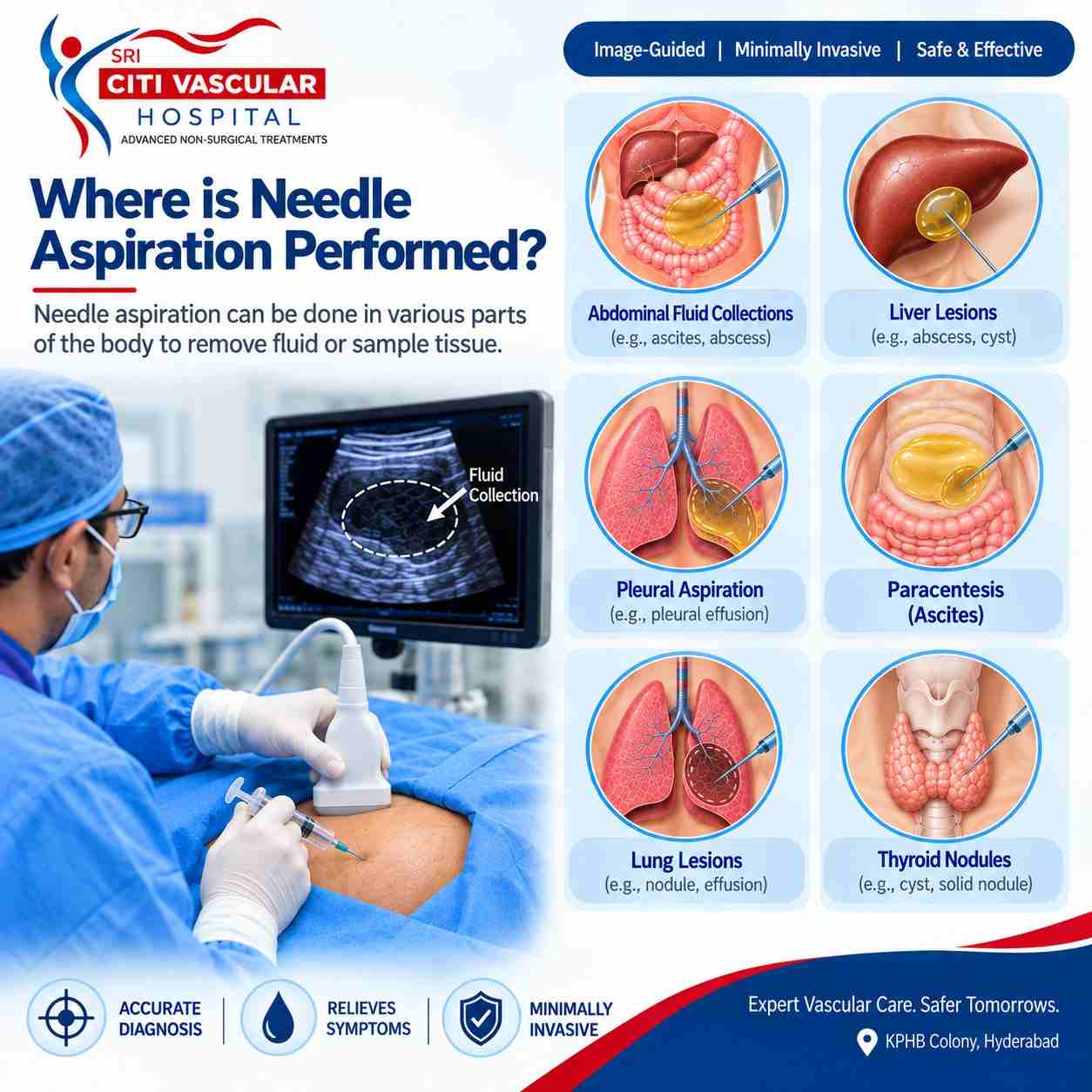
Common Needle Aspiration Procedures
|
Feature |
Diagnostic Aspiration |
Therapeutic Aspiration |
|
Primary Purpose |
Obtain fluid or cells for laboratory testing |
Remove fluid to relieve symptoms or treat infection |
|
Sample Use |
Bacterial culture, cytology, biochemistry, tumour markers |
Fluid discarded after removal; culture may be requested |
|
Volume Removed |
Small — only enough for testing (5–20mL) |
As much as safely possible — to decompress the collection |
|
Typical Use |
Solid lesions | Uncertain diagnosis | Lymph node sampling | Cystic lesion characterisation |
Abscess | Symptomatic cyst | Pleural effusion | Ascites | Haematoma |
|
Follow-up needed? |
Results guide further management plan |
Repeat aspiration or catheter if collection re-accumulates |
Not every collection requires a pigtail catheter drain — and not every collection can be adequately treated with a single aspiration. The choice depends on the size of the collection, its contents, the underlying cause, and whether it is likely to re-accumulate. The guidelines below apply generally; the final decision is made by Dr. Garge after reviewing the imaging.
|
Therapeutic Aspiration
|
Diagnostic Aspiration
|
Needle Aspiration vs Pigtail Catheter Drainage — When to Use Which
Not every collection requires a pigtail catheter drain — and not every collection can be adequately treated with a single aspiration. The choice depends on the size of the collection, its contents, the underlying cause, and whether it is likely to re-accumulate. The guidelines below apply generally; the final decision is made by Dr. Garge after reviewing the imaging.
|
Feature |
Needle Aspiration |
Pigtail Catheter Drainage |
|
Collection size |
Smaller, more liquid collections |
Larger or thick collections |
|
Tube left in body |
No — needle removed immediately |
Yes — catheter stays for days |
|
Session |
Single session |
Ongoing drainage over days |
|
Best For |
Simple abscess | Cyst | Effusion | Diagnostic sample |
Large abscess | Recurring collection | Ongoing fluid source |
|
If collection recurs |
Repeat aspiration or upgrade to catheter |
Drain repositioned or exchanged |
|
1 |
Review Imaging |
CT scan or ultrasound reviewed to plan precise needle access route — avoiding bowel, blood vessels, and other critical structures. Coagulation tests checked if required. |
|
2 |
Local Anaesthesia |
Skin and subcutaneous tissue along the needle path numbed with local anaesthetic. IV sedation given if required for patient comfort. |
|
3 |
Image-Guided Needle Entry |
Under real-time ultrasound or CT, the needle is guided precisely into the target collection. The operator monitors needle position continuously throughout. |
|
4 |
Fluid Withdrawal |
A syringe is used to aspirate fluid from the collection. Continued aspiration until the collection is evacuated or the maximum safe volume has been removed. |
|
5 |
Sample Processing |
Aspirated fluid sent to laboratory for culture and sensitivity, cytology, biochemical analysis, or specific tests as clinically indicated. |
|
6 |
Needle Removal & Recovery |
The needle is withdrawn after aspiration. A small dressing covers the entry point. Observation for 30–60 minutes. Same-day discharge in most cases. |
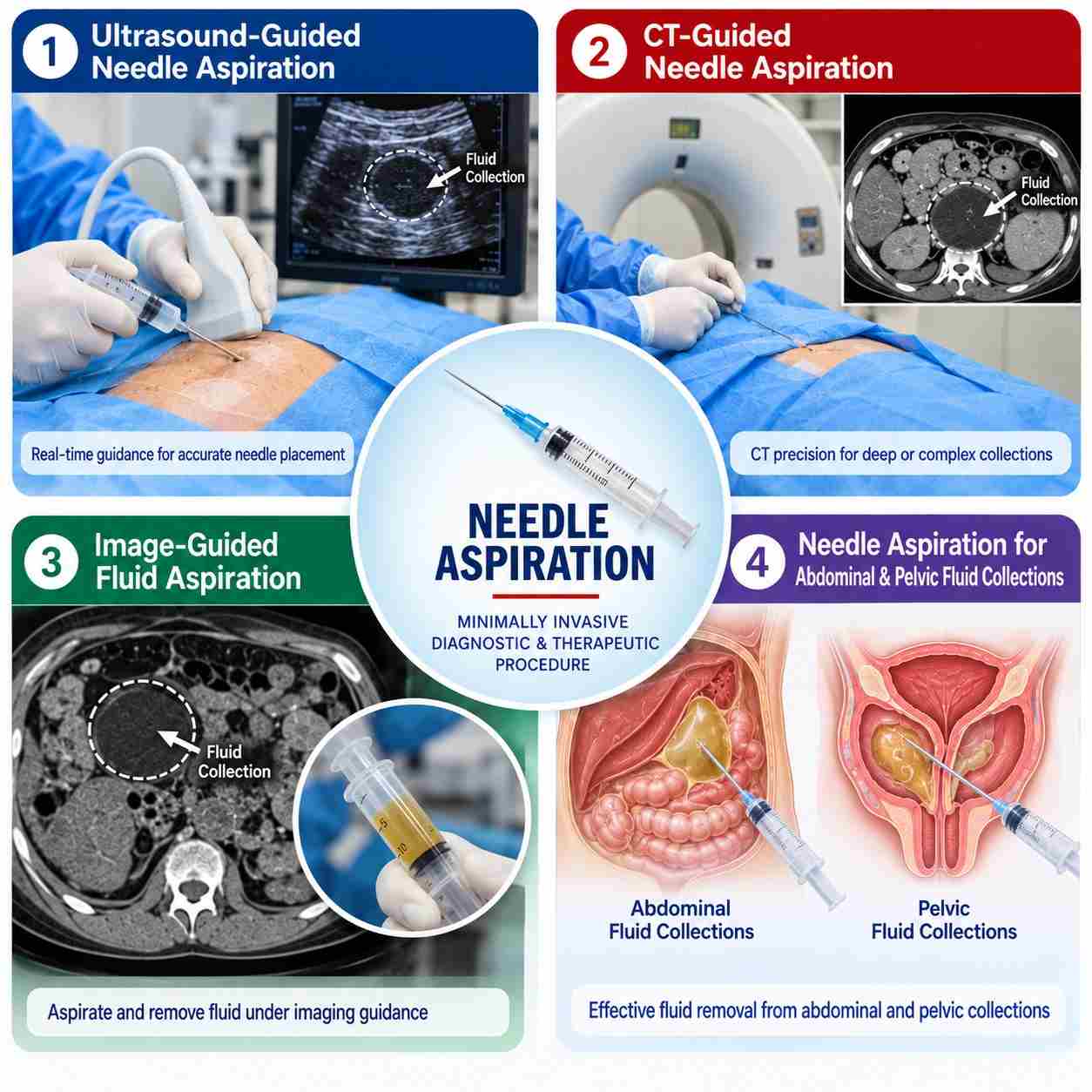
The cost of image-guided needle aspiration at Citi Vascular Centre, KPHB, Hyderabad depends on the location of the collection, the guidance modality required (ultrasound vs CT), the volume aspirated, and the laboratory tests required on the sample:
Ultrasound-guided needle aspiration — typically Rs 6,000–15,000 procedure cost
CT-guided needle aspiration (deep or complex collections) — typically Rs 8,000–20,000
Laboratory culture and sensitivity — Rs 1,000–3,000 additionally
Cytological analysis (if diagnostic aspiration) — Rs 1,000–4,000 additionally
Insurance coverage available when medically indicated — team assists with pre-auth at no extra charge
0% EMI for eligible patients | WhatsApp CT/ultrasound to 73375 83901 for an advance cost estimate