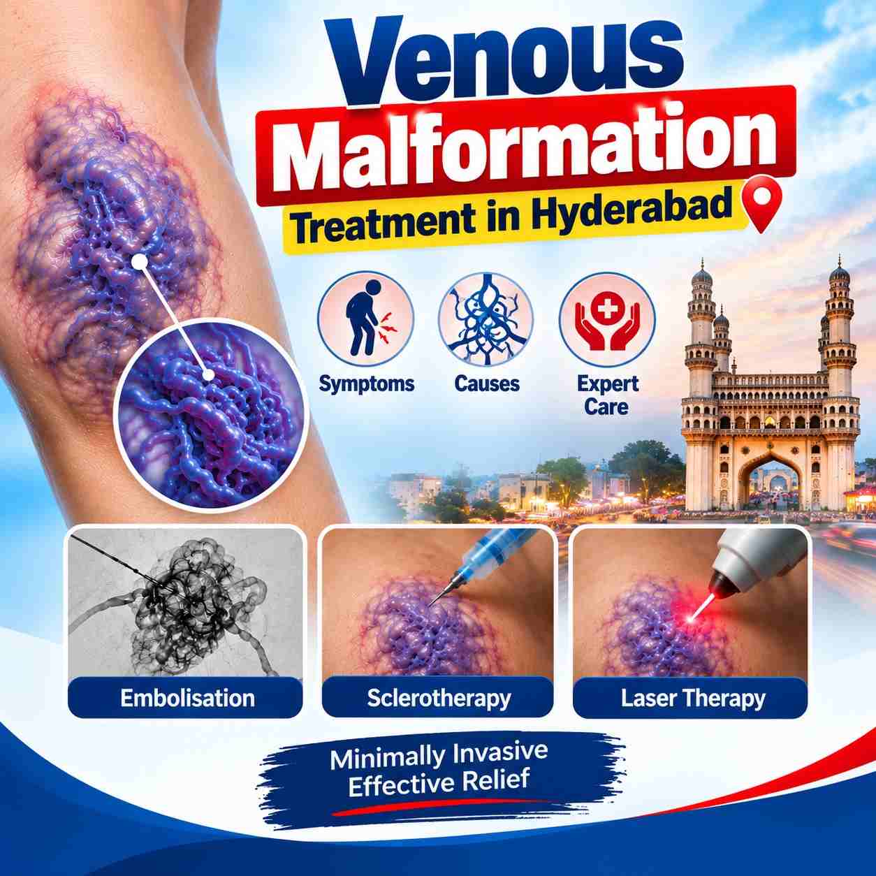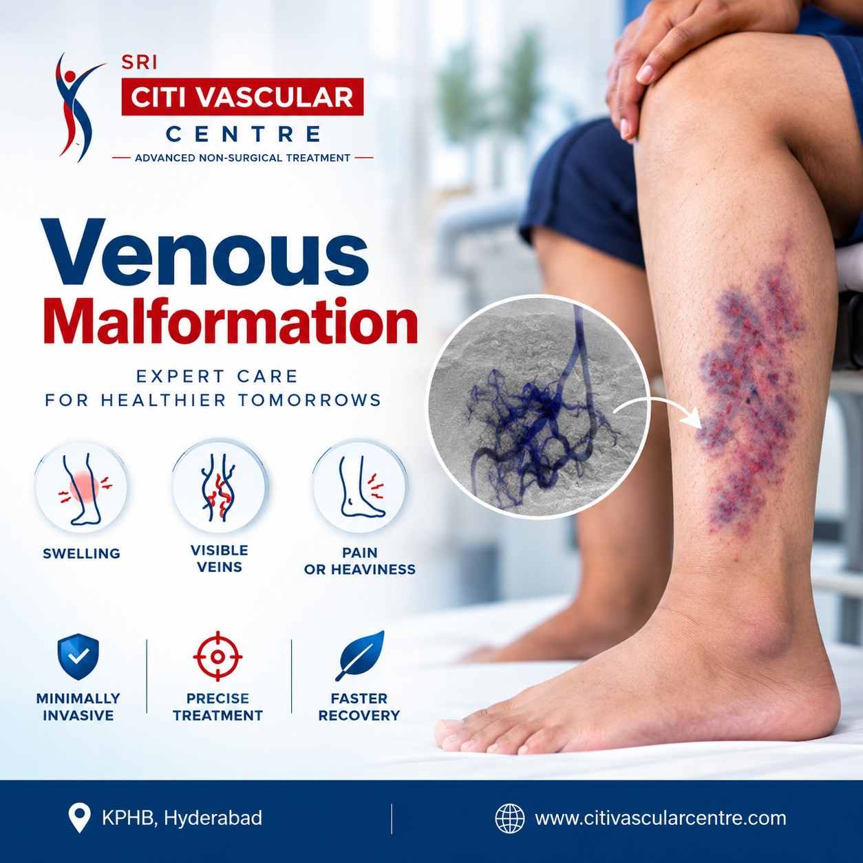
A venous malformation is a congenital, low-flow vascular malformation made up of abnormal venous channels. Venous malformation treatment depends on the lesion's size, location, depth, symptoms and involvement of nearby structures. Small, stable lesions may only need observation, while painful, enlarging or functionally significant lesions may require active treatment.
For many symptomatic venous malformations, image-guided sclerotherapy is an important minimally invasive treatment. Surgery may be appropriate for selected well-defined lesions or as part of combined treatment.
Venous malformations are usually present from birth, although they may become noticeable later.
Doppler ultrasound and MRI help establish the diagnosis and map the lesion.
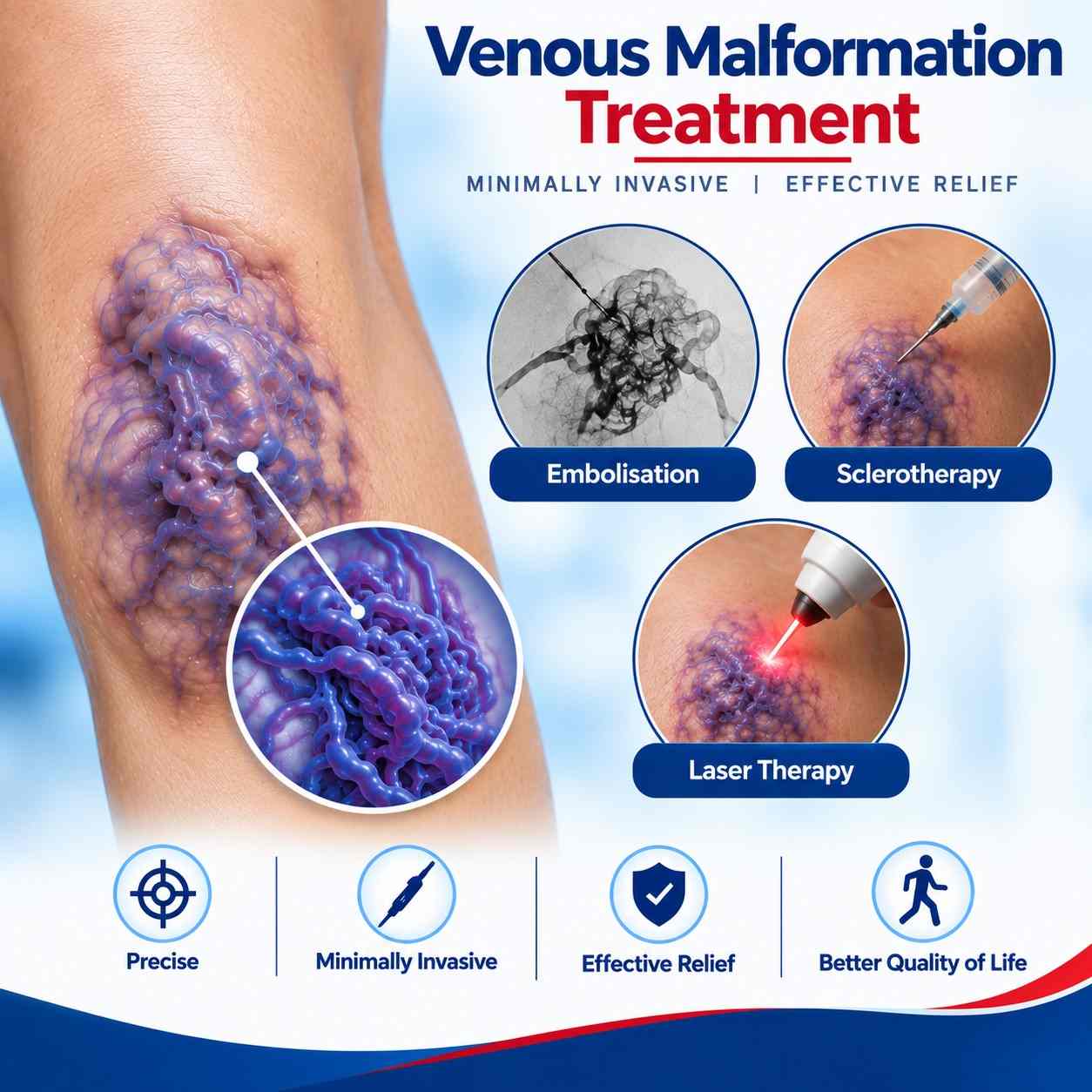
Sclerotherapy is commonly used for appropriately selected symptomatic lesions.
Foam sclerotherapy may be suitable for selected venous malformations.
Some patients need staged treatment over more than one session.
Surgery has a role in selected cases.
Treatment planning should be individualised.

Venous malformations are best evaluated by a specialist experienced in vascular anomalies and image-guided interventions, with access to appropriate imaging and multidisciplinary care when required.
At Citi Vascular Centre, KPHB, Hyderabad, Dr. Shaileshkumar Garge provides vascular and interventional radiology care, including image-guided treatment of vascular malformations.
Read more: Venous Malformation Specialist in Hyderabad
After sclerotherapy, temporary pain, swelling, tenderness or inflammation may occur. The treated area usually changes gradually rather than disappearing immediately. Recovery depends on the lesion and treatment performed.
Read more: Venous Malformation Sclerotherapy: Procedure & Recovery
Venous Malformation symptoms may vary with different stages:
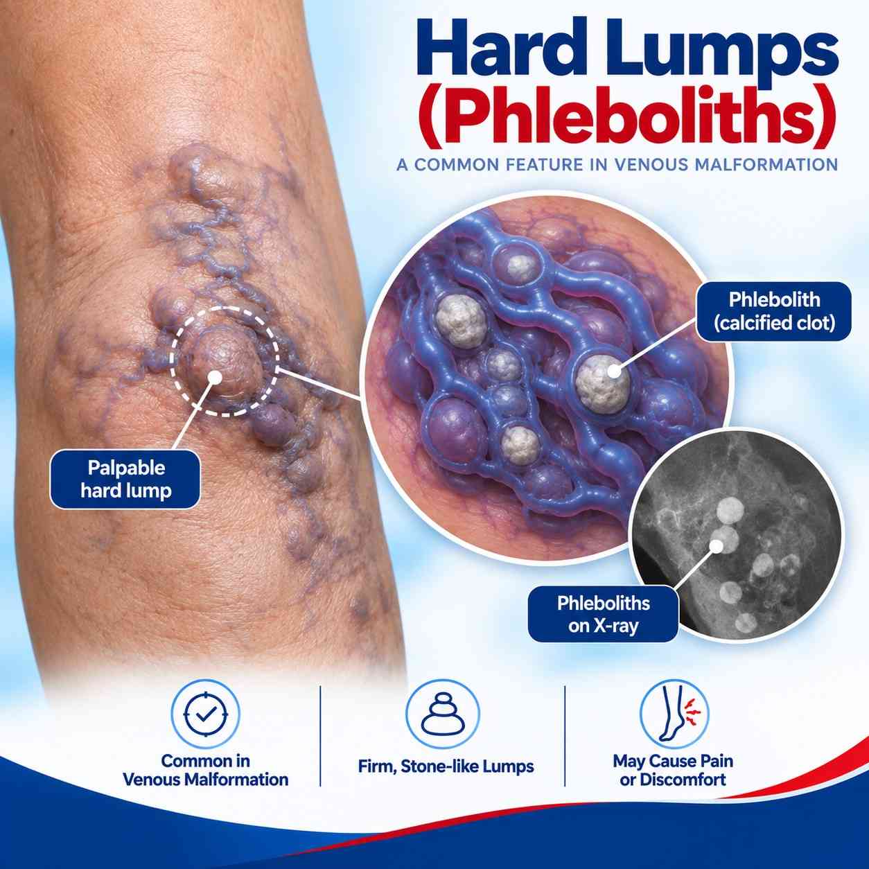
Hard Lumps (Phleboliths) Small, firm areas caused by calcified blood clots within the abnormal veins.
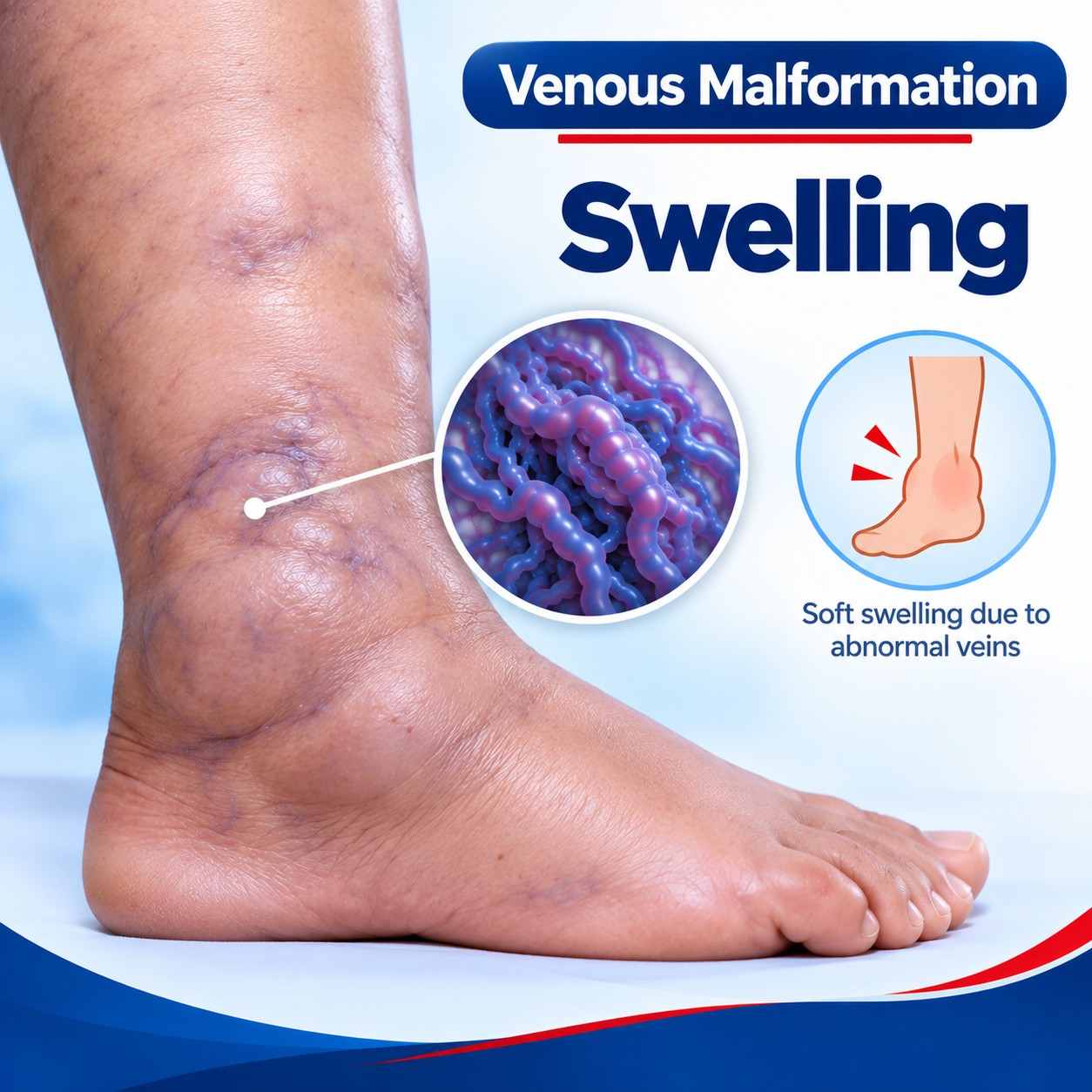
Swelling Soft swelling or a compressible lump that may become more noticeable with certain positions or activity.
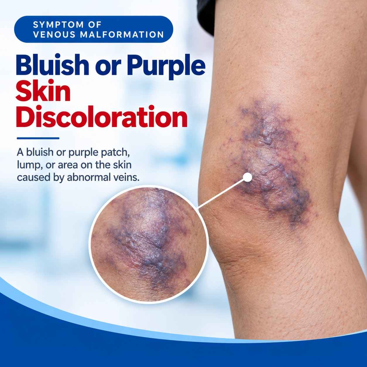
Bluish or Purple Skin Discoloration A blue or purple patch, lump, or swelling on the skin.
Diagnosis usually starts with clinical examination and Doppler ultrasound. MRI may be recommended to assess deeper tissues, the full extent of the lesion and its relationship with surrounding structures. Selected patients may also require blood tests before treatment.
Read more: Venous Malformation Diagnosis: Doppler Ultrasound & MRI
Treatment is selected according to the individual anatomy and symptoms.
Observation - Small, stable and minimally symptomatic lesions may be monitored.
Compression - Compression garments can help selected patients, particularly with limb lesions and symptoms related to venous pooling.
Sclerotherapy - Image-guided sclerotherapy is a major treatment option for symptomatic venous malformations. A sclerosant is injected into abnormal venous channels under imaging guidance, causing the treated channels to close and gradually shrink.
Foam sclerotherapy, including agents such as sodium tetradecyl sulphate (STS), may be used for appropriately selected lesions. Other sclerosants may be considered depending on the lesion.
Multiple sessions may be required.
Surgical excision may be considered when a lesion is small, localised and surgically accessible, or when residual disease remains after other treatment. Surgery is less straightforward when a malformation is extensive or infiltrates important structures.
Some complex venous malformations require a staged or combined approach, such as sclerotherapy followed by surgery for residual disease.
|
Sclerotherapy |
Surgery |
|
Minimally invasive |
Surgical removal of abnormal tissue |
|
Image-guided injection |
Excision of accessible lesion |
|
Often used for symptomatic VMs |
Selected small/localised lesions |
|
Can treat deeper components |
May provide direct removal |
|
Multiple sessions may be required |
One operation may be sufficient in selected cases |
|
Useful when surgery is difficult |
May be difficult for diffuse/infiltrating VMs |
The choice depends on anatomy, symptoms, treatment goals and involvement of surrounding structures. Neither approach is suitable for every patient.
Read more: Sclerotheraphy Vs Surgery treatments for Venous Malformation
At Citi Vascular Centre, venous malformation sclerotherapy costs approximately Rs.30,000–Rs.1,00,000 per session. A complete treatment course may cost approximately Rs. 80,000 – Rs. 3,00,000 or more, depending on the number of sessions required.
The actual cost varies with:
Size, depth and location
Imaging and procedure complexity
Sclerosant used
Anaesthesia or sedation
Number of treatment sessions
Hospital and follow-up requirements
A personalised estimate should be given after reviewing the clinical findings and imaging.
Venous malformations develop from abnormal formation of veins during development. They may appear as a bluish swelling, soft lump or compressible area and can cause pain, swelling, heaviness, tenderness or functional problems.
Read more: Venous Malformation Causes & Symptoms