
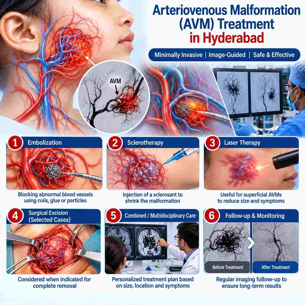
Arteriovenous malformation (AVM) treatment depends on the location, size, blood-flow pattern, feeding arteries, draining veins and symptoms. Unlike venous malformations, AVMs are high-flow vascular malformations in which arteries connect abnormally to veins.
Treatment may involve embolization, surgery, stereotactic radiosurgery or a combination of treatments, depending on the individual AVM.
AVM treatment commonly follows:
Clinical assessment → imaging → DSA angiography → treatment planning → embolization and/or surgery/radiosurgery → follow-up.
For many AVMs, embolization is used to reduce or block abnormal blood flow and may be the definitive treatment in selected lesions or part of a staged treatment plan.
AVMs are high-flow vascular malformations.
Doppler, MRI/CT and angiography may be used depending on location.
DSA is an important detailed angiographic study for treatment planning.
Embolization can be performed through a catheter without open surgery.
Liquid embolic agents such as medical glue may be used in selected AVMs.
Surgery or radiosurgery may be appropriate for selected lesions.
Complex AVMs may require staged or combined treatment.

AVM treatment requires experience in vascular anomalies, angiography and image-guided embolization, with access to appropriate imaging and surgical or radiosurgical support when required.
At Citi Vascular Centre, KPHB, Hyderabad, Dr. Shaileshkumar Garge is a vascular and interventional radiologist with FRCR (UK), FNVIR (CMC Vellore) and EBIR (Europe) qualifications. The centre provides image-guided vascular interventions and embolization procedures.
Read more: Best AVM Doctor & Hospital in Hyderabad
Most AVMs develop because blood vessels form abnormally during development. Symptoms depend on location and may include pain, swelling, warmth, pulsation, skin changes, bleeding or neurological symptoms in brain/spinal AVMs.
Diagnosis may involve Doppler ultrasound, MRI/MRA or CT/CTA. Digital subtraction angiography (DSA)provides detailed information about feeding arteries, the nidus and draining veins and is particularly important when an intervention is being planned.
Read more: AVM Causes & Symptoms | AVM Diagnosis
Treatment is individualised according to the AVM's anatomy and location.
Embolization
Endovascular embolization is a minimally invasive treatment in which a catheter is guided into the vessels supplying the AVM and an embolic material is delivered to reduce or stop abnormal blood flow.
Depending on the AVM, embolization may be used as the main treatment or before surgery/radiosurgery.
Glue Embolization
Liquid embolic agents, including medical adhesive/glue, can be delivered through a microcatheter into selected AVMs. The choice of embolic material depends on the AVM's blood-flow characteristics, vessel anatomy and treatment objective.
Other embolic agents may also be used where appropriate.
Surgery
Surgical removal may be considered for selected AVMs that can be safely accessed and completely removed. Embolization may sometimes be performed before surgery to reduce blood flow to the lesion.
Stereotactic Radiosurgery
For selected small or deep AVMs, focused radiation may be considered. The AVM closes gradually rather than being removed immediately.
Combined Treatment
Complex AVMs may require staged embolization, surgery and/or radiosurgery as part of a planned treatment strategy.
Digital Subtraction Angiography (DSA) is an X-ray-based angiographic technique that provides detailed real-time images of the AVM's feeding arteries, nidus and draining veins.
During embolization, a catheter is advanced through the blood vessels to the AVM. The interventional radiologist uses angiography to identify the abnormal vascular connections and deliver the selected embolic material.
Because AVMs can have complex vascular anatomy, treatment must be carefully planned to avoid unintended blockage of normal vessels.
|
Embolization |
Surgery |
|
Catheter-based, minimally invasive |
Open surgical procedure |
|
Uses angiographic guidance |
Direct removal of AVM |
|
Glue or other embolic agents may be used |
Requires surgical exposure |
|
May be definitive in selected AVMs |
May provide immediate removal when complete excision is feasible |
|
Can reduce blood flow before surgery |
May be preceded by embolization |
|
Often useful for difficult vascular anatomy |
Suitability depends strongly on location and accessibility |
The two approaches are not always alternatives. Embolization and surgery may be combined when clinically appropriate.
AVM treatment costs vary substantially because peripheral and brain AVMs can require very different procedures, embolic materials, hospital stays and numbers of treatment stages.
Published Indian cost sources currently place AVM treatment broadly around Rs.1.5 lakh – Rs. 5 lakh or more, while complex endovascular AVM embolization can be considerably higher depending on the anatomy and treatment plan.
Factors affecting cost include:
AVM location and size
DSA and other imaging requirements
Number of embolization sessions
Type and quantity of embolic material
Anaesthesia and procedure duration
Hospital/ICU stay
Whether surgery or radiosurgery is also required
AVM treatment costs vary substantially because peripheral and brain AVMs can require very different procedures, embolic materials, hospital stays and numbers of treatment stages.
Published Indian cost sources currently place AVM treatment broadly around ?1.5 lakh–?5 lakh or more, while complex endovascular AVM embolization can be considerably higher depending on the anatomy and treatment plan.
Factors affecting cost include:
AVM location and size
DSA and other imaging requirements
Number of embolization sessions
Type and quantity of embolic material
Anaesthesia and procedure duration
Hospital/ICU stay
Whether surgery or radiosurgery is also required
Read more: AVM Treatment Cost in Hyderabad
Recovery depends on the AVM's location, treatment performed and whether embolization was staged or combined with surgery.
After embolization, patients may have temporary discomfort or swelling at the access or treated area. Brain or spinal AVM treatment may require closer neurological observation and a longer recovery period.
Follow-up imaging is important to assess treatment response.