LAST MEDICALLY REVIEWED:
August 2026 — Dr. Shaileshkumar Garge
Citi Vascular Hospital, KPHB Colony, Road No. 1, Hyderabad, Telangana 500072
QUICK ANSWER
What Is Trigeminal Neuralgia and How Is It Treated?
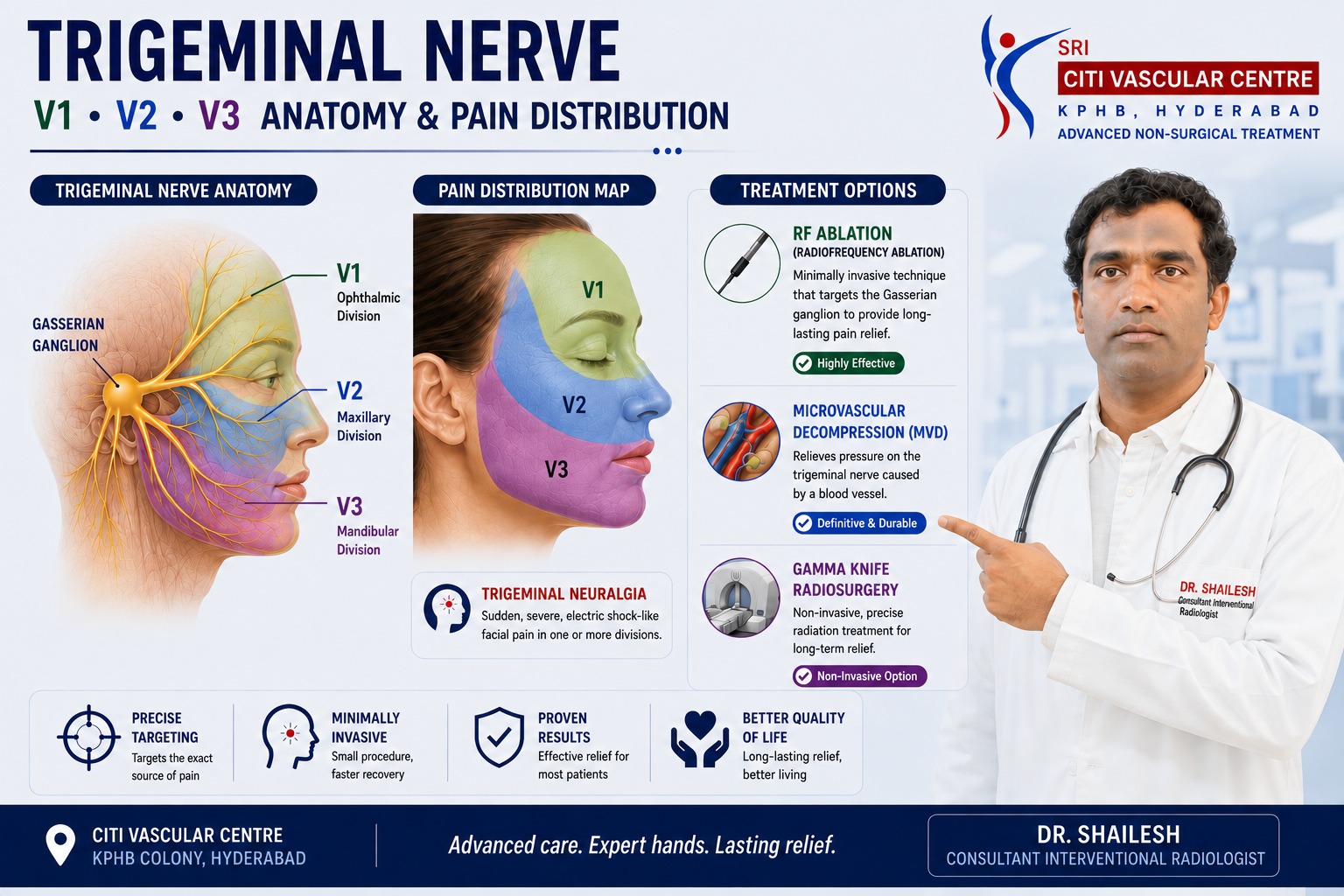
Trigeminal neuralgia is a neurological pain disorder causing sudden, severe, usually one-sided attacks of facial pain — often described as an electric shock — triggered by ordinary activities such as touching the face, eating, or speaking. The pain follows the V1 (forehead/eye), V2 (cheek/upper teeth), or V3 (jaw/chin) divisions of the trigeminal nerve. Treatment begins with medications (carbamazepine or oxcarbazepine as first-line). When medications are inadequate or poorly tolerated, RF ablation of the Gasserian ganglion, microvascular decompression, balloon compression, glycerol rhizotomy, or Gamma Knife radiosurgery can be considered based on the individual patient's profile. Dr. Garge FRCR (UK) | Citi Vascular Centre, KPHB, Hyderabad | +91-73375 83901.
Trigeminal neuralgia can cause severe, recurrent facial pain that affects eating, speaking, personal care and quality of life. Accurate diagnosis and an individualised treatment plan are important for managing the condition. A person may appear completely normal between attacks, yet live in a constant state of fear that the next electric shock through their face is only a sip of water or a brush of the teeth away. This is not ordinary facial pain. It is one of the most severe pain syndromes documented in medical literature, and it deserves both accurate diagnosis and a genuinely individualised treatment approach.
This pillar page is the comprehensive overview of trigeminal neuralgia — covering what the condition is, how V1, V2, and V3 pain patterns differ, what causes it, how it is diagnosed (and why MRI findings must be interpreted carefully), and a complete guide to all available treatments. Each treatment section includes honest discussion of what the procedure does, who it suits, and what the known limitations are. For a detailed account of any specific treatment, use the internal links to our dedicated treatment pages within this trigeminal neuralgia content cluster.
Trigeminal Neuralgia Assessment — Citi Vascular Centre, KPHB, Hyderabad
Call +91-73375 83901 | WhatsApp 73375 83901 | citivascularcentre.com | Mon–Sat 9AM–6PM
Book your Appointment here: | Appoinment: |Call +91-7032508468| Whatsapp:+91-7337583901
|
Feature |
Detail |
|
Condition Type |
Neurological pain disorder affecting the trigeminal nerve — the main sensory nerve of the face |
|
Pain Character |
Sudden, severe, electric shock-like, stabbing, or shooting — typically brief but may be repetitive throughout the day |
|
Typical Laterality |
Usually one-sided (unilateral). Bilateral TN is uncommon and should prompt investigation for secondary causes. |
|
Classic Trigger |
Light touch — washing the face, brushing teeth, chewing, talking, cold air, or even air movement against the face |
|
Nerve Divisions |
V1 (forehead/eye) | V2 (cheek/upper teeth) | V3 (jaw/chin/lower teeth) — one or more may be involved |
|
Primary Diagnosis |
Clinical — based on the characteristic pain pattern and triggers. MRI provides important supporting information. |
|
First-Line Treatment |
Carbamazepine or oxcarbazepine — medications that reduce abnormal nerve excitability |
|
Procedural Options |
RF ablation (Gasserian ganglion) | Microvascular decompression | Balloon compression | Glycerol rhizotomy | Gamma Knife |
|
Recurrence |
Possible after all treatments — recurrence rates and timing vary by procedure type and patient characteristics |
The trigeminal nerve is the fifth cranial nerve and the principal sensory nerve of the face. It transmits sensations of touch, temperature, pressure, and pain from the skin and deep structures of the face to the brainstem. It has three major branches — the ophthalmic (V1), maxillary (V2), and mandibular (V3) divisions — which together cover virtually the entire face, forehead, and upper neck. The mandibular division (V3) also carries motor fibres that control the muscles of chewing.
In trigeminal neuralgia, this nerve — or its central pathways — becomes capable of generating extremely intense, paroxysmal pain that bears no proportion to the stimulus that triggered it. A gentle touch to the cheek produces the sensation of an electric shock. The taste of food triggers a violent stabbing pain in the jaw. The act of speaking generates repeated bolts of pain across the face. This is not exaggerated sensitivity to normal pain — it is fundamentally abnormal neural activity producing pain that a healthy nervous system would not generate.
Important — not all facial pain is trigeminal neuralgia: Dental disease, sinus problems, temporomandibular disorders, migraine variants, post-herpetic neuralgia, cluster headache, and other neurological conditions can all produce facial pain. The diagnosis of trigeminal neuralgia requires a characteristic clinical pattern — recurrent, electric-shock-like, brief attacks triggered by light touch — and cannot be confirmed by imaging alone. Years of inappropriate dental treatment for undiagnosed TN are unfortunately not uncommon.
The clinical pattern of classical trigeminal neuralgia is distinctive enough that an experienced clinician can often make the diagnosis from the patient's description alone. The pain is characterised by:
Sudden onset — the pain arrives without warning, usually full-intensity from the first fraction of a second
Severe — described by sufferers as among the most intense pain they have ever experienced
Electric-shock quality — 'like lightning', 'like a live wire in my face', 'like current passing through my cheek'
Brief duration — individual attacks typically last seconds to about two minutes, though repeated attacks may feel continuous
Unilateral — almost always affecting one side of the face at a time
Triggerability — normal, harmless stimuli reliably provoke attacks in the same facial zone
Free intervals — many patients have periods with no pain between attack phases
The free intervals between attacks are one of the most clinically important diagnostic features — and also one of the cruelest aspects of the condition. A patient may appear completely well, eat a meal without incident, and then be ambushed by the next attack. The unpredictability, and the anticipatory anxiety it generates, is one of the most disabling aspects of trigeminal neuralgia.
Trigeminal neuralgia does not have a single universal cause. The underlying mechanism depends significantly on the type of TN and the individual patient's anatomy and medical history.
|
Cause |
Clinical Notes |
|
Neurovascular compression |
A blood vessel — most commonly an artery — comes into contact with the trigeminal nerve root near the brainstem (root-entry zone). In classical TN, this contact is associated with morphological nerve changes (displacement, indentation, or atrophy). Simple contact without nerve deformation is not sufficient to confirm clinically significant compression. |
|
Multiple sclerosis (MS) |
MS can damage the myelin protecting the trigeminal nerve pathways. TN in the context of MS tends to occur in younger patients and may be bilateral. Treatment considerations differ from classical TN. |
|
Tumors and structural lesions |
Meningioma, epidermoid tumors, schwannoma, and other cerebellopontine angle or skull-base lesions can compress the trigeminal nerve. This is one reason MRI is important in all patients with trigeminal neuralgia — to exclude a structural secondary cause. |
|
Other structural or inflammatory causes |
Trauma, arteriovenous malformations, post-irradiation changes, and selected inflammatory conditions affecting the trigeminal pathway can produce trigeminal pain. |
|
Idiopathic — no identifiable cause |
Some patients with a classical clinical TN pattern have no identifiable structural cause on MRI. The diagnosis is still valid if the clinical criteria are met — it is termed idiopathic TN in current classification systems. |
|
Division |
Medical Name |
Key Facial Area |
Typical TN Pain Location |
|
V1 |
Ophthalmic |
Forehead, eye, upper nose |
Forehead, eyebrow, upper eyelid, eye region, bridge of nose — electric shocks triggered by touching the brow or forehead skin |
|
V2 |
Maxillary |
Cheek, upper lip, side of nose, upper teeth |
Cheek, nasolabial fold, upper lip, upper gum, upper teeth — often initially mistaken for dental pain |
|
V3 |
Mandibular |
Lower jaw, chin, lower teeth, jaw muscles |
Lower jaw, chin, lower lip, lower teeth — V3 motor function also supplies the chewing muscles |
V2 involvement is the most commonly reported pattern in classical trigeminal neuralgia, followed by combined V2+V3. V1-only disease is the least common and requires special attention during any interventional treatment because the cornea — the transparent front surface of the eye — depends on V1 for its protective sensory innervation. More than one division can be involved simultaneously, and the pain map should be carefully documented at every assessment because treatment targeting depends on it.
|
Trigger Category |
Specific Examples |
|
Tactile / Touch |
Touching the cheek, chin, or nose | Washing or drying the face | Brushing the teeth | Shaving | Applying makeup | Clothing touching the face |
|
Oral activity |
Eating | Chewing | Swallowing | Drinking (particularly cold liquids) | Licking the lips |
|
Speech and facial movement |
Talking | Smiling | Laughing | Yawning | Opening the mouth widely |
|
Thermal / Air |
Cold air | Wind | Air conditioning | Two-wheeler riding | Exposure to cold environments |
|
Dental care |
Tooth brushing | Dental examination | Dental procedures — patients may refuse dental care because of pain fear |
|
Internal triggers |
Swallowing | Speaking | Changes in facial muscle tension |
A clinically important question to ask every patient is: 'Are there specific spots on your face that, if touched, reliably trigger an attack?' These are called trigger zones — small areas of the face or oral mucosa where light stimulation consistently provokes an attack. The presence of trigger zones is a highly characteristic feature of trigeminal neuralgia and helps distinguish it from other types of facial pain that are generally not reliably triggered by light touch.
The current International Classification of Headache Disorders (ICHD-3) and the European Academy of Neurology (EAN) guideline distinguish three main categories of trigeminal neuralgia, each with different clinical implications for diagnosis and treatment planning.
|
Type |
Definition and Clinical Features |
|
Classical Trigeminal Neuralgia |
Associated with neurovascular compression of the trigeminal nerve root that produces morphological nerve changes — such as displacement, indentation, atrophy, or flattening of the nerve root. Simple vascular contact without nerve deformation is not sufficient for this classification. A 2024 meta-analysis confirmed that these morphological changes are substantially more frequent on symptomatic than asymptomatic trigeminal nerves. |
|
Secondary Trigeminal Neuralgia |
Caused by a neurological disease or structural abnormality other than neurovascular compression — most commonly multiple sclerosis or a space-occupying lesion (meningioma, epidermoid, or other cerebellopontine angle tumor). Red flags: younger age, bilateral pain, associated neurological signs, progressive neurological deficit. |
|
Idiopathic Trigeminal Neuralgia |
Classical clinical pattern of TN without an identifiable cause on MRI. Not synonymous with psychogenic or functional pain — it is a genuine neurological diagnosis made when the clinical features are characteristic but no structural cause is identified. |
There is no blood test, nerve conduction study, or imaging finding that alone confirms trigeminal neuralgia. Diagnosis is primarily clinical — based on the characteristic pain pattern — supported by MRI and neurological examination. The clinical history is the most important diagnostic tool available.
|
Diagnostic Component |
What the Specialist Evaluates |
|
Pain quality |
Electric, shock-like, stabbing, or shooting — versus constant aching, throbbing, or burning (which are atypical and suggest an alternative or overlapping diagnosis) |
|
Duration of attacks |
Seconds to 2 minutes for individual attacks. Longer continuous pain between attacks is atypical and should prompt reconsideration of diagnosis. |
|
Trigeminal distribution |
Does the pain correspond to V1, V2, or V3 territories? Can the patient point to where the pain starts and where it travels? |
|
Triggerability |
Can the pain be reliably provoked by light touch or oral activity? Are there identifiable trigger zones? This is a highly characteristic feature of TN. |
|
Neurological examination |
Is facial sensation normal? Is the corneal reflex intact? Is jaw opening symmetric? Abnormal sensation between attacks may suggest secondary TN. |
|
Response to medication |
Carbamazepine is sufficiently specific that a positive response can support (though not confirm) the diagnosis. Lack of response may prompt diagnostic reconsideration. |
|
MRI |
Evaluates for neurovascular compression, MS plaques, tumors, and other structural causes. High-resolution T2 sequences and MR angiography are the standard protocol. MRI findings must always be interpreted in the clinical context — never in isolation. |
MRI is an important and recommended part of the trigeminal neuralgia evaluation — the EAN guideline recommends MRI when possible — but interpreting what MRI shows requires specific clinical understanding that patients should be aware of before their scan.
Vascular Contact:
A blood vessel can be seen touching or lying close to the trigeminal nerve on MRI. This is common even in people without trigeminal neuralgia. Vascular contact alone does not confirm that the vessel is causing the patient's pain and does not automatically indicate surgery.
Neurovascular Compression:
A vessel in contact with the trigeminal nerve root that also produces morphological nerve changes — displacement, indentation, flattening, or atrophy. These changes make the MRI finding clinically more meaningful. A 2024 meta-analysis confirmed these changes are significantly more frequent on symptomatic nerves.
The practical implication for patients: 'A blood vessel is touching my trigeminal nerve on MRI' is not the same as 'I have neurovascular compression causing my trigeminal neuralgia.' MRI findings are one piece of clinical information that must be interpreted alongside the pain history, neurological examination, and the clinical diagnosis of TN. A specialist who recommends surgery solely on the basis of vascular contact — without matching clinical features of TN and without evidence of nerve deformation — is not following current international guidelines.
Book your Appointment here: | Appoinment: |Call +91-7032508468| Whatsapp:+91-7337583901
For most patients, treatment begins with medication. Medicines reduce the frequency and severity of attacks by suppressing abnormal nerve excitability. The decision about which medication, at what dose, and for how long should always be made by a physician with knowledge of the patient's comorbidities, current medications, and individual risk factors.
|
Medication |
EAN Status |
Clinical Notes |
|
Carbamazepine |
First-line (Level A evidence) |
The most established medication for classical TN. Highly effective initially. Requires blood tests for monitoring. Side effects include dizziness, unsteadiness, cognitive effects, skin reactions. HLA-B*1502 testing recommended before starting in South Asian patients due to SJS risk. |
|
Oxcarbazepine |
First-line |
Similar mechanism to carbamazepine with a somewhat different side-effect profile. Also considered a first-line option in current guidelines. Generally requires similar monitoring. |
|
Lamotrigine |
Second-line |
Used as add-on treatment or when first-line agents are not tolerated. Slow dose titration required to minimise skin reaction risk. |
|
Gabapentin / Pregabalin |
Supporting evidence |
Used in selected patients — particularly in secondary TN or when first-line agents are ineffective or poorly tolerated. Evidence base for TN is less robust than for other pain conditions. |
|
Baclofen |
Supporting evidence |
May be used alone or in combination with carbamazepine. Particularly considered in patients with multiple sclerosis-associated TN. |
|
Botulinum Toxin Type A |
Emerging evidence |
Injected into trigger zones in selected patients. Evidence growing but not yet first-line. |
Do not stop trigeminal neuralgia medication suddenly without medical advice. Some of these medications require gradual dose reduction. If your medication is no longer controlling the pain, causing unacceptable side effects, or needs adjustment — discuss this with your treating physician rather than stopping independently. Call +91-73375 83901.
Medication no longer being adequate is the point at which most patients are referred for procedural or surgical treatment. The common reasons for this are: pain breaks through despite adequate doses; the dose required for control causes unacceptable sedation or cognitive effects; the attacks are becoming more frequent; or the patient is unable to eat, drink, or maintain oral hygiene because of pain. When this situation arises, the six procedural options below should be discussed with the patient in a structured way.
Radiofrequency ablation of the Gasserian ganglion is a minimally invasive, fluoroscopy-guided procedure in which a specialised RF electrode is positioned through the foramen ovale (a natural opening in the skull base) toward the trigeminal ganglion. Electrical stimulation identifies the specific V1, V2, or V3 division that is causing the patient's pain before controlled radiofrequency heat is applied to interrupt the abnormal pain transmission. The procedure is performed under local anaesthesia and IV sedation and typically produces rapid pain relief.
Key clinical points: The procedure is neuroablative — it intentionally disrupts nerve fibres — and facial numbness is a common expected outcome. Pain can recur as the nerve recovers, and long-term recurrence rates increase with follow-up duration. V1 involvement requires specific caution about corneal sensation. Repeat procedures are possible. RF is particularly valuable when the patient is not a suitable candidate for open surgery, when MRI does not show convincing neurovascular compression that MVD could address, or when rapid relief from a minimally invasive approach is the priority.
For the complete, detailed guide to Gasserian ganglion RF ablation — including the 7-step procedure, sensory stimulation, energy delivery, corneal risk, risk table, and full FAQ — see our dedicated page: Radiofrequency Ablation of the Gasserian Ganglion for Trigeminal Neuralgia [internal link to /gasserian-ganglion-radiofrequency-ablation-trigeminal-neuralgia].
Microvascular decompression is fundamentally different from all other treatments for trigeminal neuralgia in one important way: it is the only option that attempts to address the underlying cause rather than deliberately damaging the nerve to reduce pain. During MVD, a neurosurgeon opens the skull behind the ear (retrosigmoid craniotomy), accesses the area where the trigeminal nerve root meets the brainstem, and places a small protective material between the nerve and the offending blood vessel — separating them to relieve the compression.
Because MVD does not intentionally injure the trigeminal nerve, it offers the possibility of meaningful pain relief without the deliberate sensory trade-off of neuroablative procedures. Published evidence and the EAN guideline support MVD as the preferred first surgical option for classical TN with convincing neurovascular compression in patients who are suitable candidates for intracranial surgery. Long-term pain-free rates after MVD are generally higher than after percutaneous procedures in comparative studies.
The important limitation is that MVD is an intracranial operation with surgical and anaesthetic risks that are substantially different from percutaneous treatments — including the small but real risks associated with craniotomy: cerebellar injury, cerebrospinal fluid leak, hearing loss, facial weakness, infection, and stroke. Patients must be medically fit for general anaesthesia and intracranial surgery. MVD is not appropriate for every patient — and for patients without convincing neurovascular compression, its theoretical rationale is weaker.
Percutaneous balloon compression is another minimally invasive option performed through the foramen ovale — the same access route as RF ablation. A thin catheter with a small inflatable balloon at its tip is guided toward the trigeminal ganglion under fluoroscopic imaging. The balloon is inflated for a defined period to mechanically compress the trigeminal ganglion, selectively injuring the pain-transmitting fibres while producing a predictable degree of facial numbness and, often, temporary chewing weakness (because the motor fibres of V3 run through the ganglion).
Balloon compression has a particular advantage over RF in one specific clinical situation: it does not require the patient to communicate stimulation responses during the procedure, meaning it can be performed under full general anaesthesia. This makes it a practical option for patients who cannot cooperate with the stimulation phase of RF. Comparative published evidence — including a 2026 systematic review and meta-analysis — found broadly comparable outcomes between RF and balloon compression across several major endpoints, though the authors appropriately noted that this does not establish formal equivalence.
Glycerol rhizotomy is a percutaneous chemical procedure that uses the same foramen ovale access route. Contrast material is used to confirm positioning within the trigeminal cistern (the CSF-filled space containing the trigeminal nerve root and ganglion), and then glycerol — a chemical that selectively damages myelin — is injected to produce a controlled neurolytic effect on the pain-transmitting fibres. No balloon is inflated and no radiofrequency energy is used.
Glycerol rhizotomy can provide meaningful pain relief and has been used for trigeminal neuralgia for decades. Potential advantages include the absence of radiofrequency energy delivery and the fact that stimulation confirmation is not required. Limitations include somewhat variable selectivity — it is more difficult to direct glycerol to a specific trigeminal division than to confirm division-specific targeting by stimulation during RF — and published comparative reviews suggest recurrence rates with glycerol may be somewhat higher than with RF or MVD in some patient populations. Sensory changes and facial numbness are still possible outcomes.
Stereotactic radiosurgery — most commonly known by the brand name Gamma Knife — delivers highly focused radiation to the trigeminal nerve root using external beams precisely convergent on the target from multiple directions. Unlike RF, balloon compression, and glycerol rhizotomy, Gamma Knife treatment requires no needle entering the skull base and no open surgery. The radiation is delivered during a single outpatient session under local anaesthetic to secure the frame — there are no incisions and no anaesthesia in the traditional sense.
The most clinically important difference from all other procedures is the delayed onset of effect. Gamma Knife treatment does not produce immediate pain relief — most patients require weeks to months before experiencing the full therapeutic response. This makes it unsuitable for patients who need urgent pain relief. It is particularly useful for patients who prefer a non-incisional approach, who are medically unsuitable for other procedures, or who have previously undergone other treatments. Facial numbness can develop over time after radiosurgery, and recurrence is possible. Long-term follow-up in published series shows recurrence rates that increase over years.
|
Feature |
RF Ablation |
MVD |
Balloon |
Glycerol |
Gamma Knife |
|
Approach |
Percutaneous |
Open craniotomy |
Percutaneous |
Percutaneous |
Non-incisional radiation |
|
Nerve deliberately injured |
Yes |
No |
Yes |
Yes |
Yes |
|
Immediate pain relief |
Usually rapid |
Usually rapid |
Usually rapid |
Usually rapid |
Delayed — weeks/months |
|
Facial numbness |
Common |
Less common |
Common |
Possible |
Possible — delayed |
|
Treats compression cause |
No |
Yes — if present |
No |
No |
No |
|
Suitable for V1 |
Caution — cornea |
Yes |
Yes — motor V3 risk |
Less selective |
Yes |
|
Major surgical risk |
Low |
Intracranial — significant |
Low |
Low |
Radiation dose planning |
|
Can be repeated |
Usually yes |
More complex |
Usually yes |
Usually yes |
Possible |
No single treatment is best for every patient. The choice depends on the trigeminal division involved, MRI findings (particularly whether convincing neurovascular compression is present), the patient's age and surgical fitness, previous treatments, the speed of pain relief needed, the patient's preference regarding facial numbness, and the available local expertise. At Citi Vascular Centre, KPHB, Dr. Garge discusses all applicable options honestly before any treatment is recommended. Call +91-73375 83901.
Many patients with trigeminal neuralgia wait too long before seeking specialist assessment — sometimes years of unnecessary dental procedures, ENT investigations, or inadequate pain management pass before the correct diagnosis is made. The following are the most important clinical signals that specialist assessment is indicated:
Facial pain that is brief, electric-shock-like, or stabbing rather than constant or aching
Pain reliably triggered by light touch, eating, speaking, or cold air — particularly in a reproducible facial zone
Pain consistently on one side of the face only — though bilateral TN does exist
Pain repeatedly attributed to teeth or sinuses without dental or sinus pathology being found
Medication that worked initially but is no longer providing adequate control
Medication causing unacceptable side effects that limit the dose that can be used
Pain preventing eating, drinking, or maintaining oral hygiene
Facial numbness, bilateral symptoms, or other neurological findings alongside facial pain — investigate for secondary causes
Q1: What is trigeminal neuralgia?
A: Trigeminal neuralgia is a neurological pain disorder affecting the trigeminal nerve — the main sensory nerve of the face. It causes sudden, severe, electric shock-like or stabbing attacks of facial pain, typically on one side, triggered by ordinary activities such as touching the face, eating, speaking, or cold air. The V1, V2, and V3 divisions of the nerve may be involved.
Q2: What causes trigeminal neuralgia?
A: The most common identifiable cause is neurovascular compression — a blood vessel in contact with the trigeminal nerve root that causes morphological nerve changes (displacement, indentation, or atrophy). Other causes include multiple sclerosis, tumours, and structural lesions. Some patients have classical TN with no identifiable structural cause. Simple vascular contact on MRI without nerve deformation is not sufficient to confirm clinically significant compression.
Q3: Why does trigeminal neuralgia feel like an electric shock?
A: The electric quality reflects abnormal neural discharge in the trigeminal system — rather than graded sensory transmission, the nerve produces sudden high-amplitude bursts of activity in response to normally innocuous stimuli. This is similar in principle to a short circuit in an electrical system, generating an amplified signal from a minimal input.
Q4: Which branch causes cheek and upper teeth pain?
A: Cheek pain, nasolabial area pain, upper lip pain, and upper teeth pain most commonly correspond to V2 — the maxillary division. V2 disease is one of the most common presentations of trigeminal neuralgia. Patients are often initially assessed by a dentist, and the absence of dental disease despite severe tooth-area pain should raise the possibility of TN.
Q5: Is MRI necessary for trigeminal neuralgia?
A: Yes — MRI is an important part of the TN evaluation and is recommended by the EAN guideline when possible. It investigates for neurovascular compression (with morphological nerve changes), multiple sclerosis, tumours, and other structural causes. However, MRI alone does not establish the diagnosis — TN is primarily a clinical diagnosis based on the characteristic pain pattern.
Q6: Does a blood vessel touching the trigeminal nerve mean I need surgery?
A: Not automatically. Vascular contact with the trigeminal nerve is common even in people without TN. Clinically meaningful neurovascular compression requires morphological nerve changes — displacement, indentation, atrophy, or distortion — in addition to vascular contact. The MRI finding must always be interpreted alongside the clinical diagnosis. Surgery based on vascular contact alone, without matching TN clinical features and nerve deformation, is not supported by current guidelines.
Q7: What is the first treatment for trigeminal neuralgia?
A: Carbamazepine or oxcarbazepine are the recommended first-line medications according to major guidelines including the EAN. Both reduce abnormal nerve excitability. They require medical monitoring and may cause side effects. When medication fails to control pain adequately or causes unacceptable adverse effects, procedural or surgical treatment is discussed.
Q8: When is RF ablation considered for trigeminal neuralgia?
A: RF ablation of the Gasserian ganglion is considered when medication does not provide adequate control, when the patient prefers a minimally invasive approach over open surgery, when MRI does not show convincing neurovascular compression that MVD could address, or when the patient is not a suitable candidate for craniotomy. It can also be considered when pain recurs after previous treatment.
Q9: Is microvascular decompression better than RF ablation?
A: For classical TN with convincing neurovascular compression and a patient suitable for intracranial surgery, MVD is generally the preferred surgical option — it addresses the underlying cause without intentionally ablating the nerve, and has higher long-term pain-free rates in comparative studies. For patients without convincing compression, who cannot undergo surgery, or who prefer a percutaneous approach, RF and other percutaneous procedures are appropriate alternatives.
Q10: What is balloon compression for trigeminal neuralgia?
A: Balloon compression is a percutaneous procedure in which a small balloon catheter is passed through the foramen ovale to the Gasserian ganglion and briefly inflated to mechanically compress and selectively injure pain-transmitting fibres. It can be performed under general anaesthesia without requiring the patient to report stimulation responses, making it suitable when RF stimulation is not feasible.
Q11: What is glycerol rhizotomy?
A: Glycerol rhizotomy is a percutaneous chemical procedure in which glycerol — a neurolytic agent — is injected into the trigeminal cistern via the foramen ovale. It selectively damages pain-transmitting myelin fibres. No radiofrequency energy or balloon is used. It can provide meaningful pain relief, though it is less division-specific than RF and some comparative reviews report somewhat higher recurrence rates.
Q12: What is Gamma Knife treatment for trigeminal neuralgia?
A: Gamma Knife is stereotactic radiosurgery — focused radiation aimed at the trigeminal nerve root without surgery or needle placement. The key clinical difference is delayed onset: most patients require weeks to months before experiencing full pain relief. It is useful for patients who prefer a non-incisional approach or cannot undergo other procedures, but is not appropriate when urgent pain relief is needed.
Q13: Can trigeminal neuralgia affect both sides of the face?
A: Bilateral TN is uncommon — approximately 1–4% of TN cases. When it occurs, secondary causes should be investigated carefully, particularly multiple sclerosis. Bilateral TN at presentation — rather than successive unilateral episodes on different sides over years — is a red flag that warrants thorough neurological assessment and MRI.
Q14: Can trigeminal neuralgia come back after treatment?
A: Yes — recurrence is possible after all treatments for TN. MVD generally has the best long-term pain-free rates among surgical options. Percutaneous procedures (RF, balloon, glycerol) and Gamma Knife are all associated with increasing recurrence rates at longer follow-up. Recurrence does not mean the original treatment failed — it often reflects natural nerve recovery after neuroablation or disease progression over time
Q15: Is trigeminal neuralgia curable?
A: Some patients achieve very long-lasting remission — particularly after successful MVD for classical TN with convincing neurovascular compression. However, recurrence is possible after all treatments, and 'cure' is not a term that can be reliably applied. Long-term pain control and remission are more accurate ways to describe the realistic treatment goals.
Q16: What are the red flags in trigeminal neuralgia that need investigation?
A: Red flags requiring additional investigation: bilateral pain at presentation | persistent facial numbness between attacks | significant sensory loss | jaw weakness not explained by V3 motor involvement | hearing changes | visual symptoms | multiple cranial nerve abnormalities | younger than typical presentation | history of multiple sclerosis or cancer | progressive neurological deficit. These suggest secondary TN and require thorough assessment.
Q17: Can V1 trigeminal neuralgia be treated with RF?
A: Yes, but with specific additional caution. V1 (ophthalmic division) supplies sensation to the cornea. Significant reduction in corneal sensation from RF ablation can impair the protective blink reflex, potentially leading to corneal dryness, exposure, ulceration, and vision loss. Any patient with V1 involvement being considered for RF requires careful ophthalmological awareness and thorough informed consent about corneal risk.
Q18: What happens after trigeminal neuralgia treatment?
A: Follow-up should assess pain relief, facial sensation quality (numbness, tingling, dysesthesia), trigger sensitivity, ability to eat and speak normally, jaw strength (V3), corneal sensation (V1), and medication requirements. A meaningful outcome is not just 'is the pain gone' but 'can the patient eat, brush their teeth, wash their face, and speak without fear of triggering an attack.'
Q19: What questions should I ask before TN treatment?
A: Important questions: Which trigeminal division is my pain in? Does my MRI show neurovascular compression with nerve deformation, or just vascular contact? Why is this specific treatment recommended for me? How much facial numbness should I expect? What is the risk to my cornea? How long does pain relief typically last? What happens if pain returns? Can this treatment be repeated? What are the alternatives?
Q20: Who performs trigeminal neuralgia RF ablation in Hyderabad?
A: Dr. Shaileshkumar Garge — FRCR (UK), FNVIR (CMC Vellore), EBIR (Spain) — Director and Chief Vascular Physician at Citi Vascular Centre, KPHB Colony, Hyderabad, performs Gasserian ganglion radiofrequency ablation as part of comprehensive trigeminal neuralgia care, including MRI review, clinical diagnosis confirmation, and honest discussion of all treatment options before any procedure is planned. Call +91-73375 83901.
|
Reference |
Key Relevance |
|
Bendtsen L, Zakrzewska JM et al. EAN Guideline on Trigeminal Neuralgia. Eur J Neurol. 2019;26:831–849. |
Primary evidence-based guideline: carbamazepine/oxcarbazepine first-line; MVD recommended for classical TN when surgery appropriate; surgery when medication inadequate or poorly tolerated. |
|
ICHD-3 (International Classification of Headache Disorders, 3rd Edition). |
Defines classical TN requiring neurovascular compression with morphological nerve changes — not simple vascular contact. Establishes diagnostic criteria for all TN subtypes. |
|
Meta-analysis of MRI morphological changes in TN. 2024. |
Confirmed that nerve atrophy, displacement, flattening, indentation, and distortion are substantially more frequent on symptomatic trigeminal nerves — supporting the morphological-changes criterion for classical TN. |
|
Systematic reviews of percutaneous TN treatments — RF, balloon, glycerol. |
Multiple systematic reviews confirm all three percutaneous neuroablative procedures provide meaningful pain relief with different recurrence and complication profiles. None is universally superior. |
|
RF vs balloon compression systematic review and meta-analysis. 2026. |
Found broadly comparable outcomes between RF thermocoagulation and percutaneous balloon compression across several major endpoints — cautions that comparable outcomes should not be interpreted as formal equivalence. |
Trigeminal neuralgia is one of the most severe pain syndromes in medicine — and one of the most treatable when correctly diagnosed and appropriately managed. The characteristic electric-shock attacks, triggered by ordinary touch or facial activity in the V1, V2, or V3 trigeminal territories, create a clinical picture that is distinctive enough for an experienced specialist to recognise from the clinical history. MRI provides valuable supporting information — but MRI vascular contact without nerve deformation is not sufficient to confirm classical TN or to recommend surgery.
Treatment follows a logical progression: medication first (carbamazepine or oxcarbazepine are the EAN-endorsed first-line agents), then procedural treatment when medication is inadequate or poorly tolerated. The procedural options — RF ablation of the Gasserian ganglion, microvascular decompression, balloon compression, glycerol rhizotomy, and Gamma Knife radiosurgery — each have specific advantages, limitations, and complication profiles that make them appropriate for different patient subgroups. No single procedure is the best for every patient. The right treatment is the one that provides the best balance of pain relief, durability, neurological safety, and quality of life for the individual.
At Citi Vascular Centre, KPHB Colony, Hyderabad, Dr. Shaileshkumar Garge provides a comprehensive trigeminal neuralgia assessment — including MRI review, clinical diagnosis confirmation, and honest discussion of all applicable treatment options — before any procedure is planned. For a detailed account of Gasserian ganglion RF ablation, see our dedicated page [internal link]. Call +91-73375 83901 or WhatsApp 73375 83901.
Trigeminal Neuralgia — Expert Assessment at Citi Vascular Centre, KPHB, Hyderabad
MRI Review | Clinical Diagnosis | RF Ablation | MVD Coordination | All Options Discussed
Dr. Shaileshkumar Garge | FRCR (UK) | FNVIR (CMC Vellore) | EBIR (Spain) | 12+ Years | 15,000+ Procedures
Call +91-73375 83901 | WhatsApp 73375 83901 | citivascularcentre.com
KPHB Colony, Hyderabad | Mon–Sat 9AM–6PM | Outstation Welcome
Book your Appointment here: | Appoinment: |Call +91-7032508468| Whatsapp:+91-7337583901