LAST MEDICALLY REVIEWED:
November 2026 — Dr. Shaileshkumar Garge
Citi Vascular Hospital, KPHB Colony, Road No. 1, Hyderabad, Telangana 500072
1. Quick Answer — What Is an IVC Filter?
2. What Is the Inferior Vena Cava (IVC)?
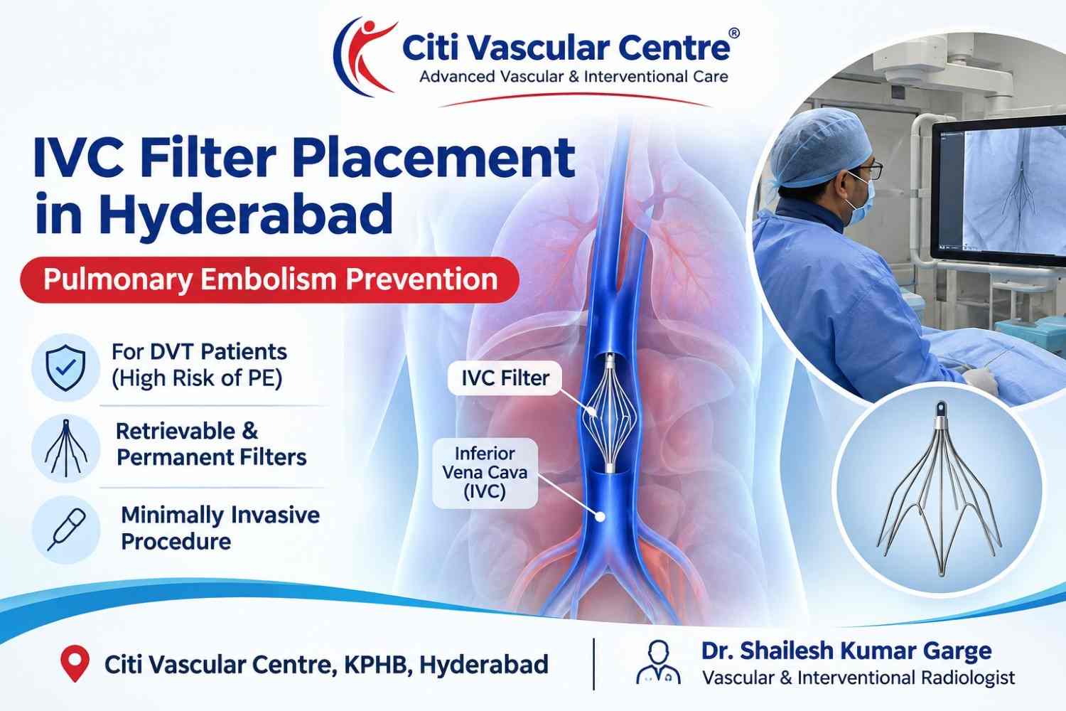
3. What Is an IVC Filter and How Does It Work?
5. Retrievable vs Permanent IVC Filters — Which Is Better?
6. How Is IVC Filter Placement Performed? (Step-by-Step)
7. What Happens After IVC Filter Placement?
8. IVC Filter and Anticoagulation — Are Both Needed?
9. Risks and Complications of IVC Filter Placement
10. IVC Filter Removal — Can It Be Taken Out?
11. Cost of IVC Filter Placement in Hyderabad
12. Why Choose Citi Vascular Centre?
13. Dr. Shailesh Kumar Garge — IVC Filter Specialist, Hyderabad
14. Frequently Asked Questions
The inferior vena cava is the largest vein in the human body. It runs vertically along the right side of the spine, collecting oxygen-depleted blood from the legs, pelvis, and abdominal organs and carrying it back to the right side of the heart.
Why does this matter for DVT? When a blood clot forms in a deep vein of the leg — a condition called deep vein thrombosis — there is always a risk that the clot or a piece of it will break off, travel up through the IVC, and lodge in the pulmonary arteries of the lung. This is called a pulmonary embolism (PE), and it can be life-threatening.
An IVC filter works by sitting inside this large vein like a net, catching migrating clots before they can reach the heart and lungs.
Pulmonary embolism is responsible for approximately 100,000–180,000 deaths per year in the United States alone. In India, the true burden is significantly underdiagnosed. DVT and PE together are known as venous thromboembolism (VTE).
An IVC filter is a small, metallic, cone-shaped or umbrella-shaped device — typically about 2–3 cm in diameter when deployed. It is collapsed into a thin delivery system for insertion and then springs open inside the IVC, anchoring itself to the vein wall with small hooks or struts.
Once in place, the filter's wire struts form a mesh that traps large clots (thromboemboli) travelling upward through the IVC. Blood continues to flow normally around and through the filter. The trapped clot is gradually broken down by the body's own clot-dissolving system over days to weeks.
What an IVC Filter Does and Does Not Do
|
An IVC filter DOES: |
An IVC filter DOES NOT: |
|
Catch migrating blood clots before they reach the lungs |
Treat or dissolve the original DVT clot in the leg |
|
Reduce the immediate risk of fatal pulmonary embolism |
Replace anticoagulation (blood-thinning) treatment |
|
Allow time for safer anticoagulation to begin or resume |
Prevent DVT from recurring in the legs |
|
Protect during thrombolysis when large clots are being dissolved |
Guarantee complete prevention of all PE (small clots may pass through) |
Not every patient with DVT needs an IVC filter. It is recommended in specific clinical situations where the risk of pulmonary embolism is high and anticoagulation alone is insufficient or unsafe.
Definite (Absolute) Indications
Anticoagulation is contraindicated — recent major surgery, active internal bleeding, intracranial haemorrhage, haemorrhagic stroke, or severe thrombocytopenia (very low platelet count)
Anticoagulation has failed — DVT or PE occurred despite adequate blood-thinning treatment
Complication of anticoagulation — the patient developed serious bleeding while on blood thinners
Relative (Conditional) Indications
Large, free-floating thrombus in the iliac vein or IVC — high risk of breaking off
Massive pulmonary embolism already occurred — prevents a second potentially fatal embolism
Poor cardiopulmonary reserve — the patient cannot survive another PE even if small
Before catheter-directed thrombolysis (CDT) — during active clot dissolution, fragments may break off; a filter provides protection during the procedure
High-risk surgical patients — major orthopaedic or cancer surgery when anticoagulation must be held
Non-compliant patients who are unlikely to take anticoagulants reliably
⚠️ Important: IVC filters are not a treatment for DVT — they are a protection against PE. The underlying DVT must still be treated with anticoagulation, catheter-directed thrombolysis, or thrombectomy as clinically appropriate.
This is one of the most important decisions in IVC filter placement. Historically, all IVC filters were permanent — once placed, they stayed forever. Modern filters are retrievable, meaning they can be removed once the risk period has passed.
|
Feature |
Retrievable Filter |
Permanent Filter |
|
Design |
Hooks/struts designed for both anchoring and later retrieval |
Fixed anchoring mechanism — not designed for removal |
|
Removal possible? |
Yes — typically within 3–6 months (some up to 12 months) |
No — remains in the body permanently |
|
Best for |
Most DVT patients — temporary PE protection while anticoagulation is contraindicated or during CDT |
Patients with lifelong contraindication to anticoagulation; very limited indications today |
|
Long-term risks |
Lower — if retrieved promptly; can convert to permanent if retrieval fails |
Higher — filter fracture, migration, IVC perforation, IVC thrombosis over years |
|
Cost |
Slightly higher — retrieval is a separate procedure |
One-time cost |
|
Current preference |
Strongly preferred by interventional radiologists globally |
Reserved for selected cases only |
At Citi Vascular Centre, we place retrievable filters in the vast majority of cases and plan for retrieval at the time of placement. Studies show that up to 70% of IVC filters placed globally are never retrieved — this is a clinical problem, as long-term indwelling filters carry significant complication risks. We actively follow up all our filter patients to ensure timely removal.
IVC filter placement is a short, minimally invasive endovascular procedure performed in a catheterisation laboratory (cath lab) under real-time X-ray (fluoroscopy) guidance. Here is exactly what happens:
|
Step |
Stage |
What Happens |
|
1 |
Pre-procedure preparation |
Blood tests (clotting profile, kidney function), imaging review (CT or Doppler to confirm IVC anatomy and clot extent). The procedure site (groin or neck) is cleaned and draped. IV access is established. |
|
2 |
Local anaesthesia |
Local anaesthetic is injected at the access site. Most patients do not require general anaesthesia or sedation, though mild sedation can be offered for anxious patients. The procedure is essentially painless. |
|
3 |
Venous access |
A thin needle punctures a vein — most commonly the right internal jugular vein (neck) or the right common femoral vein (groin). A guidewire is advanced into the IVC under fluoroscopy. |
|
4 |
IVC venogram |
Contrast dye is injected to map the IVC — measuring its diameter, identifying the renal veins (the filter must be placed below them), and checking for any anomalies such as a double IVC or renal vein variants. |
|
5 |
Filter deployment |
The filter is loaded into a thin delivery catheter (typically 7–12 French). It is advanced to the correct position just below the renal veins and released. The filter springs open and anchors to the IVC wall. |
|
6 |
Confirmation imaging |
A final venogram or fluoroscopic check confirms the filter is correctly positioned, fully expanded, and anchored. The delivery system is removed and a small pressure dressing is applied to the access site. |
|
7 |
Post-procedure monitoring |
The patient is observed for 2–4 hours. Access site is checked for bleeding or haematoma. Most patients are discharged the same day or the following morning. |
Duration: 30–60 minutes | Anaesthesia: Local anaesthesia (± mild sedation) | Hospital stay: Day procedure / overnight
The jugular (neck) approach is preferred when there is a large DVT in the femoral or iliac vein, as it avoids passing through the clot-bearing vessel and reduces the risk of dislodging the thrombus during insertion.
Recovery from IVC filter placement is straightforward for most patients. The procedure itself is so minimally invasive that many patients are surprised by how quickly they feel back to normal.
Immediately after the procedure
The access site (groin or neck) is monitored for 2–4 hours for bruising or bleeding
Blood pressure, heart rate, and oxygen levels are checked regularly
The patient can eat and drink normally unless sedation was used
Most patients walk to the ward and are discharged within hours
At home — first week
Mild bruising or tenderness at the access site is normal — this resolves within 5–7 days
Avoid strenuous lifting or vigorous exercise for 48–72 hours
A light dressing is kept over the access site for 24 hours
Blood-thinning medication (anticoagulation) will usually be restarted or continued — this is essential to treat the underlying DVT even with the filter in place
Ongoing management
A follow-up appointment is scheduled — typically at 4–6 weeks — to reassess the DVT and plan filter retrieval
The patient carries a filter card identifying the filter type, lot number, and date of placement — important if medical imaging is needed in the future
Most IVC filters are MRI-compatible, but the treating team should be informed before any MRI scan
Retrieval should be planned before 3–6 months if a retrievable filter was placed.
This is a question patients frequently ask — if I have a filter, do I still need blood thinners?
The answer, in most cases, is yes — and this is an important distinction to understand.
|
Scenario |
What This Means |
|
Filter placed because anticoagulation is TEMPORARILY contraindicated (e.g. recent surgery) |
Anticoagulation should be restarted as soon as it is safe to do so. The filter provides short-term protection in the meantime. Once anticoagulation is established and the PE risk decreases, the filter should be retrieved. |
|
Filter placed before catheter-directed thrombolysis (CDT) |
Anticoagulation continues throughout the CDT procedure. The filter protects against PE while the clot is being actively dissolved and fragments may break off. Filter is retrieved after CDT is complete. |
|
Permanent filter placed due to lifelong anticoagulation contraindication |
Blood thinners may not be possible long-term. The filter provides ongoing protection. Regular follow-up is needed to monitor for filter complications and IVC thrombosis. |
The key point: an IVC filter treats the risk of PE but does not treat the DVT. The clot in your leg or pelvis still needs to be managed — either with anticoagulation alone or with catheter-directed clot removal
IVC filter placement is a safe, well-established procedure with a low complication rate. However, like any medical intervention, it carries some risks — both procedure-related and long-term.
Procedure-Related Risks (Short-Term)
|
Complication |
Frequency |
Notes |
|
Access-site haematoma |
2–4% |
Bruising at the groin or neck; resolves spontaneously |
|
Contrast reaction |
< 1% |
Mild rash to severe anaphylaxis (very rare); pre-medication given in high-risk patients |
|
Air embolism |
Very rare |
Prevented by careful technique; head-down positioning during insertion |
|
Filter malposition |
< 2% |
Pre-deployment IVC venogram minimises malpositioning risk |
|
Arterial puncture |
< 1% |
Ultrasound-guided access significantly reduces this risk |
Long-Term Risks (Permanent or Long-Dwelling Filters)
|
Long-Term Complication |
What Happens |
How to Reduce Risk |
|
IVC thrombosis |
Clot forming on the filter body, blocking the IVC itself |
Anticoagulation; timely filter retrieval |
|
Filter migration |
Filter moves from its original position — may travel toward the heart |
Correct sizing at deployment; retrieval before strut fatigue |
|
Filter fracture |
Metal struts break off and can migrate to the heart or lungs |
Retrieve before 12 months if possible |
|
IVC perforation |
Filter struts penetrate through the IVC wall — usually asymptomatic |
More common with long dwell time; early retrieval preferred |
|
Failed retrieval |
After long dwell time, filter struts embed deeply making removal difficult |
Plan retrieval within 3–6 months; specialised retrieval techniques available |
Key safety message: The long-term complication rate of retrievable IVC filters increases significantly the longer they remain in place. If a retrievable filter was placed and anticoagulation has been safely restarted, the filter should be removed — ideally within 3–6 months.
Studies show that filters left in place for more than 12 months have significantly higher rates of fracture, perforation, and thrombosis. This is why filter retrieval planning starts at the time of placement at Citi Vascular Centre.
Yes — retrievable filters can be removed once the risk of pulmonary embolism has decreased to an acceptable level. Filter removal is a separate endovascular procedure, typically performed under local anaesthesia via a jugular vein approach. A snare catheter grasps the filter's retrieval hook and collapses it into a sheath for removal.
When Is Filter Retrieval Appropriate?
Anticoagulation has been safely established or the contraindication has resolved
The acute PE risk period has passed (typically 3–6 months)
The underlying DVT has been treated with thrombolysis or is stable on anticoagulation
The filter has not been in place so long that strut embedding makes removal unsafe
What If the Filter Cannot Be Removed?
In cases where the filter has been in place for a long time and standard retrieval is difficult, specialised advanced retrieval techniques — using laser sheaths, forceps, or endobronchial forceps — can often achieve removal even in challenging cases. These are performed by experienced interventional radiologists.
The cost of IVC filter placement varies depending on the type of filter used, the access approach, and whether the procedure is performed as an emergency or elective case.
|
Procedure |
Approximate Cost (Hyderabad) |
|
IVC Filter Placement (retrievable) |
₹1,00,000 – ₹2,00,000 |
|
IVC Filter Placement (permanent) |
₹80,000 – ₹1,50,000 |
|
IVC Filter Removal (retrieval procedure) |
₹60,000 – ₹1,20,000 |
|
IVC Filter + CDT (combined — DVT thrombolysis + filter) |
₹2,50,000 – ₹5,00,000 |
What is included in the cost at Citi Vascular Centre:
• Pre-procedure imaging review and clinical consultation
• IVC filter device (filter cost is a major component — brand and type vary)
• Procedure in a dedicated cath lab with fluoroscopy
• Radiological contrast and consumables
• Post-procedure monitoring and discharge planning
• IVC filter patient card and retrieval planning appointment
Insurance: IVC filter placement is covered under most cashless health insurance policies as a cardiovascular / vascular intervention. Pre-authorisation is required. Contact our billing team with your policy details and treating doctor's referral for documentation assistance.
|
✅ |
Dedicated Cath Lab |
Full digital angiography suite with fluoroscopy and ultrasound — the standard of care for IVC filter procedures |
|
✅ |
Retrieval-First Philosophy |
Every retrievable filter placed with a documented retrieval plan — not placed and forgotten |
|
✅ |
Emergency Filter Placement |
Available when urgently required for high-risk PE patients — no delays waiting for OT scheduling |
|
✅ |
Advanced Retrieval Capability |
Specialised techniques for difficult filter retrievals — late presentations, embedded struts |
|
✅ |
Ultrasound-Guided Access |
Vascular access performed under real-time ultrasound to minimise access-site complications |
|
✅ |
Coordinated DVT Care |
IVC filter placement integrated with catheter-directed thrombolysis and venous stenting — full DVT treatment under one roof |
|
✅ |
Transparent Pricing |
All-inclusive costs quoted upfront; insurance documentation support provided |
Dr. Shailesh Kumar Garge is the Director and Chief Vascular & Interventional Radiologist at Citi Vascular Centre, KPHB Colony, Hyderabad.
15+ years of experience in interventional radiology and vascular access procedures
Specialist in acute DVT management — catheter-directed thrombolysis, IVC filters, venous stenting
Expert in IVC filter retrieval including complex late-presentation cases
Image-guided procedures performed in a fully equipped cath lab with real-time fluoroscopy
Consultations available in Telugu, Hindi, and English
Affiliated with Citi Vascular Centre, KPHB Colony — easily accessible from Kukatpally, Miyapur, Balanagar, Kompally, and central Hyderabad
Book a Consultation — IVC Filter Placement, Hyderabad
Citi Vascular Centre | KPHB Colony, Hyderabad — 500072
call: +91 73375 83901
www.citivascular.com
Dr. Shailesh Kumar Garge | Senior Interventional Radiologist
Bring: DVT imaging report (Doppler / CT) · Current medication list · Insurance card
⚠️ For sudden chest pain, breathlessness, or rapid heartbeat — this may be a pulmonary embolism. Go to the nearest emergency department immediately
1Q: What is an IVC filter and why is it placed?
A: An IVC filter is a small cage-like device placed inside the inferior vena cava — the large vein carrying blood from the legs to the heart. It catches blood clots that break off from deep vein thrombosis (DVT) in the legs before they can travel to the lungs and cause a potentially fatal pulmonary embolism (PE). It is typically recommended when blood-thinning treatment is not safe or has not worked.
2Q: Is IVC filter placement a painful procedure?
A: No. The procedure is performed under local anaesthesia at the access site — either the groin or the neck. Most patients feel only mild pressure during the procedure. It is not painful, and many patients are surprised by how quickly it is over. Mild bruising at the access site is normal and resolves within a few days.
3Q: How long does IVC filter placement take?
A: The procedure typically takes 30–60 minutes in the catheterisation laboratory. After 2–4 hours of post-procedure observation, most patients are discharged on the same day or the following morning. There is no need for general anaesthesia or a prolonged hospital stay in most cases.
4Q: Can an IVC filter be removed after placement?
A: Yes — retrievable IVC filters are designed to be removed once the risk of pulmonary embolism has decreased. Removal is typically planned at 3–6 months after placement, once anticoagulation is established and the DVT is stable or resolved. The removal procedure (filter retrieval) is a separate endovascular procedure and also takes about 30–60 minutes. We strongly recommend removal rather than leaving retrievable filters permanently in place.
5Q: Do I still need blood thinners if I have an IVC filter?
A: In most cases, yes. An IVC filter protects against pulmonary embolism but does not treat the DVT itself — the blood clot in your leg still needs treatment. When anticoagulation becomes safe again (which is usually the reason the filter was placed in the first place), it should be restarted as soon as possible. Anticoagulation also reduces the risk of new clot forming on the filter.
6Q: Is an IVC filter permanent?
A: Not necessarily. Modern retrievable filters are designed to be removed and should be removed once they are no longer needed. However, some patients — those with a lifelong contraindication to anticoagulation — may require a permanent filter. Even so, permanent filters today are reserved for a narrow group of patients. The trend in modern practice is strongly toward retrievable filters with planned removal.
7Q: What is the difference between a jugular and femoral approach for IVC filter placement?
A: The jugular (neck) approach is used when there is a large DVT in the femoral or iliac veins — accessing through the groin would mean passing instruments through or near the clot, which risks dislodging it. The jugular approach comes from above, completely avoiding the clot. The femoral (groin) approach is used when the DVT is confined to the calf or when no significant clot is present in the access vein. Both approaches deliver the same filter to the same position in the IVC.
8Q: What is the cost of IVC filter placement in Hyderabad?
A: At Citi Vascular Centre, Hyderabad, IVC filter placement (retrievable) typically costs between ₹1,00,000 and ₹2,00,000 as an all-inclusive package covering the filter device, cath lab procedure, imaging, and post-procedure care. Filter retrieval is a separate procedure costing approximately ₹60,000–₹1,20,000. Most cashless insurance policies cover this procedure. Contact us on +91 73375 83901 for a detailed cost estimate based on your case.
9Q: Can an IVC filter cause problems if left in too long?
A: Yes — this is a clinically important concern. Long-term indwelling retrievable filters are associated with increased risks of filter fracture (broken struts migrating to the heart), filter migration, IVC perforation, and IVC thrombosis (clotting of the filter itself, blocking the vena cava). These risks increase the longer the filter is left in place, particularly beyond 6–12 months. This is why timely retrieval planning is essential and why we schedule follow-up for every patient at the time of filter placement.
10Q: Who is the best doctor for IVC filter placement in Hyderabad?
A: Dr. Shailesh Kumar Garge at Citi Vascular Centre, KPHB Colony, Hyderabad is a senior Interventional Radiologist with over 15 years of experience in vascular intervention, including IVC filter placement and retrieval, catheter-directed thrombolysis, and May-Thurner syndrome treatment. His practice uses image-guided techniques in a dedicated catheterisation laboratory, and all filter patients receive a retrieval plan at the time of placement.
✅ An IVC filter catches migrating blood clots before they reach the lungs — it does not treat the DVT itself.
✅ It is recommended when anticoagulation is contraindicated, has failed, or before catheter-directed thrombolysis.
✅ Retrievable filters are strongly preferred over permanent filters in most patients.
✅ The procedure takes 30–60 minutes under local anaesthesia — most patients go home the same day or next morning.
✅ Filter retrieval should be planned at the time of placement and performed within 3–6 months.
✅ Long-term indwelling filters carry significant complication risks — fracture, migration, IVC thrombosis.
✅ Blood-thinning treatment (anticoagulation) must continue alongside the filter to treat the underlying DVT.
✅ Cost at Citi Vascular Centre: ₹1,00,000–₹2,00,000 (placement); ₹60,000–₹1,20,000 (retrieval). Insurance covered.
|
# |
Reference |
|
1 |
PREPIC Study Group. Eight-year follow-up of patients with permanent vena cava filters in the prevention of pulmonary embolism. Circulation 2005;112:416–22. — The landmark randomised trial establishing the efficacy and long-term outcomes of IVC filters. |
|
2 |
Kaufman JA, et al. Guidelines for the use of retrievable and convertible vena cava filters: report from the Society of Interventional Radiology multidisciplinary consensus conference. JVIR 2006. — Defines indications for retrievable filters and retrieval protocols. |
|
3 |
Angel LF, et al. Systematic review of the use of retrievable inferior vena cava filters. JVIR 2011. — Demonstrates increased complication rates with prolonged filter dwell time; supports early retrieval. |
|
4 |
Mismetti P, et al. (PREPIC2). Effect of a retrievable inferior vena cava filter plus anticoagulation vs anticoagulation alone on risk of recurrent pulmonary embolism. JAMA 2015;313:1627–35. — Key RCT evaluating short-term retrievable filters in PE patients. |