LAST MEDICALLY REVIEWED:
August 2026 — Dr. Shaileshkumar Garge
Citi Vascular Hospital, KPHB Colony, Road No. 1, Hyderabad, Telangana 500072
QUICK ANSWER
Osteoid Osteoma: Is Surgery or Ablation the Better Treatment?
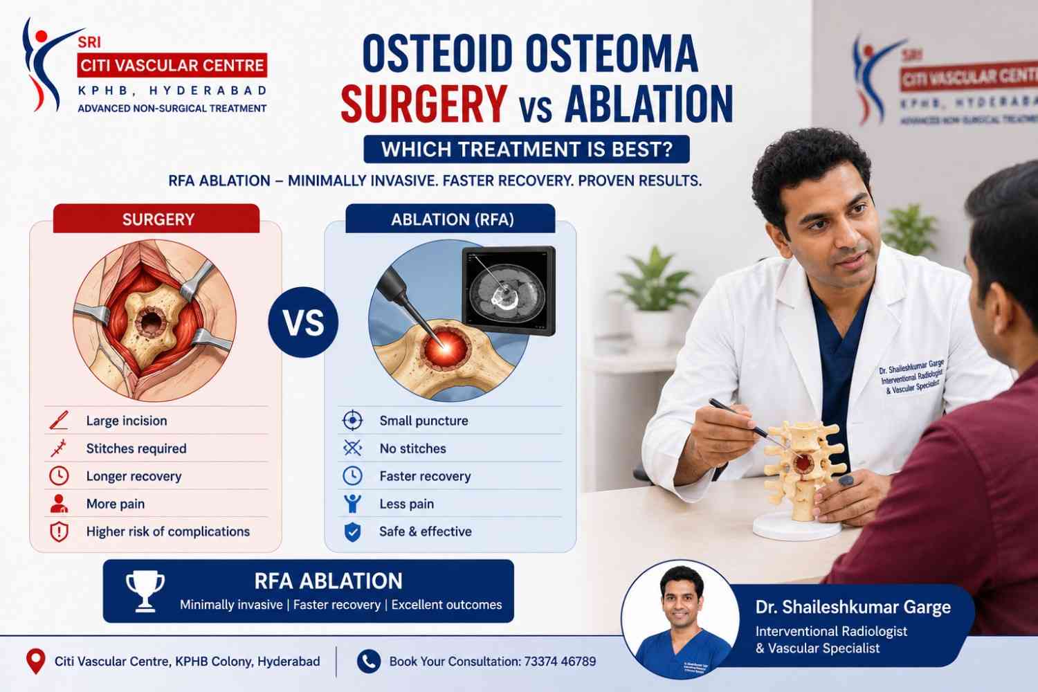
For most confirmed, accessible osteoid osteomas, CT-guided thermal ablation (RFA or microwave) is the preferred first-line treatment over surgery. It destroys the nidus precisely without removing surrounding bone, requires no open incision, offers faster recovery, and has equivalent or better clinical success rates than surgery in published systematic reviews. Surgery is still the right choice when the diagnosis is uncertain, when percutaneous access is anatomically impossible, or when other clinical factors favour an open approach. Dr. Garge FRCR (UK) | Citi Vascular Centre, KPHB, Hyderabad | +91-73375 83901.
When a patient or parent is told that an osteoid osteoma needs treatment, two paths are typically presented: surgery — specifically open surgical excision or arthroscopic resection of the nidus — and image-guided thermal ablation, most commonly CT-guided radiofrequency ablation (RFA) or microwave ablation. Both treatments work by eliminating the nidus, the small central core of the osteoid osteoma that is responsible for its characteristic severe pain. But they do so in fundamentally different ways, with meaningfully different implications for scarring, recovery, bone preservation, and the risk of recurrence.
This comparison page addresses the question patients most commonly ask — 'Which is better for me?' — with an honest, evidence-based answer that considers the clinical evidence, the specific advantages of each approach, and the specific situations where one is clearly preferable to the other. For the step-by-step procedure details of ablation, see our Osteoid Osteoma Ablation Procedure page. For RFA-specific evidence data, see our Osteoid Osteoma RFA page.
Discuss Your Options — Citi Vascular Centre, KPHB, Hyderabad
Call +91-73375 83901 | WhatsApp 73375 83901 | citivascularcentre.com | Mon–Sat 9AM–6PM
|
Feature |
CT-Guided Thermal Ablation |
Open / Arthroscopic Surgery |
|
Approach |
Percutaneous needle through skin and bone to nidus under CT |
Surgical incision, bone drilling or resection of nidus + margin |
|
Incision |
None — tiny needle entry only |
Surgical wound requiring closure |
|
Bone Removed |
Nidus only — surrounding bone preserved |
Nidus + safety margin of surrounding bone |
|
Anaesthesia |
GA (children, complex) or conscious sedation (adults) |
General anaesthesia — always |
|
Hospital Stay |
Same-day or overnight discharge |
1–3 days ward admission in most cases |
|
Primary Success Rate |
91–96% (systematic review 3,023 patients) |
85–95% (varies by technique and margin adequacy) |
|
Recurrence Rate |
8.3% — repeat ablation effective in most |
Varies widely — curettage without margin: higher | En bloc resection: lower but more bone removed |
|
Scar |
None — tiny needle mark fades to invisible |
Surgical scar of variable size — depends on location |
|
Return to School / Desk Work |
5–7 days |
2–3 weeks |
|
Return to Sports — Lower Limb |
4–6 weeks (weight-bearing restriction) |
6–12 weeks — wound + bone healing |
|
Can Be Repeated? |
Yes — safely, with similar technique |
Repeat surgery is technically harder — scar tissue |
|
Diagnosis Confirmed? |
Should be confirmed — if uncertain, biopsy first |
Provides tissue specimen for definitive histology |
CT-Guided Thermal Ablation:
A specialised probe is placed into the nidus through a percutaneous needle track under CT guidance. Radiofrequency or microwave energy heats the nidus to temperatures sufficient to cause irreversible cell death — destroying the pain source without removing bone. Tissue is ablated in place, not excised. No incision. No bone resection beyond the nidus. For the complete 10-step procedure guide, see our Osteoid Osteoma Ablation Procedure page.
Open or Arthroscopic Surgery:
Open surgery involves a skin incision, exposure of the affected bone, and drilling or curettage to remove the nidus and a margin of surrounding bone. Arthroscopic approaches are used for intra-articular lesions — particularly those within the hip joint — where direct visualisation without full open access is achievable. Surgery provides a tissue specimen for histopathological confirmation but removes more bone than ablation.:
Open surgery involves a skin incision, exposure of the affected bone, and drilling or curettage to remove the nidus and a margin of surrounding bone. Arthroscopic approaches are used for intra-articular lesions — particularly those within the hip joint — where direct visualisation without full open access is achievable. Surgery provides a tissue specimen for histopathological confirmation but removes more bone than ablation.
Ablation is not limited to small lesions — it is the preferred first-line approach for most osteoid osteomas that meet basic selection criteria. The following characteristics predict a good outcome with ablation and typically favour it over primary surgery.
|
Osteoid Osteoma Characteristic |
Ablation Response |
Why Ablation Is Preferred |
|
Confirmed diagnosis on CT — lucent nidus with sclerosis |
Excellent |
Ablation targets a precisely defined target. CT-confirmed nidus is the best predictor of successful ablation. |
|
Cortical or subcortical nidus in long bone (femur, tibia, humerus) |
Excellent — standard indication |
Most common clinical scenario. Well-studied in published systematic reviews. Ablation is the established standard of care. |
|
Nidus at safe distance from skin and neural structures |
Excellent |
Thermal margin between ablation zone and skin or nerve provides a safe buffer. Standard ablation without protective measures. |
|
Nidus close to a nerve — with hydrodissection feasible |
Good — with additional precautions |
Hydrodissection displaces nerve away from ablation zone. Cryoablation may be preferred. Requires experienced operator but achieves good outcomes. |
|
Intra-articular nidus — within the hip joint |
Good — cryoablation often preferred |
Heat-based ablation near articular cartilage carries chondral injury risk. Cryoablation's cold ablation zone is more cartilage-tolerant. Arthroscopic surgery is the surgical alternative. |
|
Spinal osteoid osteoma — posterior arch, facet |
Good with experienced IR — cryoablation preferred |
Thermal protection of spinal cord and nerve roots is critical. Cryoablation allows a safer ablation zone near neural structures than heat-based techniques in selected cases. |
|
Recurrent osteoid osteoma after prior ablation |
Safe and effective — repeat ablation |
Repeat ablation is technically feasible and follows the same procedure. Published data shows secondary failure rate of only 3.1% after repeat RFA. |
|
Recovery Aspect |
Thermal Ablation |
Surgical Excision |
|
Day of Procedure |
1–3 hours recovery then home for most patients |
Ward admission — IV fluids, wound drain (sometimes), monitored overnight in most cases |
|
Pain — First 48 Hours |
Moderate procedure-site aching — paracetamol + NSAID adequate |
Surgical wound pain — stronger analgesics needed initially | Drain site discomfort if drain placed |
|
Wound Care |
None — tiny needle mark covered by adhesive strip. No wound to clean or dress. |
Wound dressing changes. Suture or staple removal at Day 10–14. Surgical wound care instructions. |
|
School / Desk Work |
Day 5–7 for most patients |
Week 2–3 — wound comfort and energy levels permitting |
|
Driving |
Day 3–5 (if lower limb not involved) |
Week 2–3 — wound healing and analgesia requirements |
|
Light Activity (walking, swimming) |
Week 2–3 (non-weight-bearing sites sooner) |
Week 3–4 — wound protection and bone healing |
|
Sports — Weight-Bearing Bone |
Week 4–6 (weight-bearing restriction during healing) |
Month 2–3 — bone healing + surgical wound + muscle recovery after open exposure |
|
Sports — Non-Weight-Bearing |
Week 2–3 |
Week 3–4 |
|
Pain Resolution — Osteoid Osteoma Night Pain |
24–72 hours after successful ablation for most patients |
Days to weeks — wound pain initially masks osteoid osteoma pain improvement |
|
Risk / Complication |
Ablation Risk Profile |
Surgery Risk Profile |
|
Wound infection |
Rare — < 1%. No wound. Skin entry seals within 24 hours. |
1–3% surgical site infection. Wound requiring antibiotic treatment and sometimes surgical debridement. |
|
Skin injury or burn |
Skin burn — reported with superficial nidus + heat-based ablation. Managed with wound dressing. Uncommon with experienced operators. |
Surgical wound dehiscence. Hypertrophic scar. Keloid formation at incision site. |
|
Nerve injury |
Risk adjacent to named nerve — mitigated by hydrodissection, cryoablation selection, and careful planning. Reported in < 1–2% with experienced operators. |
Intraoperative nerve injury during surgical dissection — risk depends on nidus proximity to nerve. Can be more difficult to predict and control than during planned ablation. |
|
Bone fracture |
Rare — more relevant for weight-bearing location with dense reactive sclerosis. Weight-bearing restriction post-procedure is the primary preventive measure. |
Bone weakening after nidus curettage — higher risk when a larger bone margin is removed. Post-operative weight-bearing restriction also required. |
|
Blood loss |
Negligible — no bone resection |
Blood loss during bone drilling and curettage — variable, rarely requiring transfusion but adds to procedural risk |
|
Anaesthetic risk |
Conscious sedation for selected adults — lower risk. GA when required — same risk as surgery. |
General anaesthesia always — carries intubation and cardiovascular risk. Relevant for patients with comorbidities. |
|
Incomplete treatment |
8.3% primary failure rate (systematic review) — repeat ablation effective in most cases |
Recurrence varies by surgical technique — curettage without adequate margin: higher | En bloc resection with margin: lower but more bone removed |
|
Incomplete tissue diagnosis |
No tissue removed — if diagnosis was uncertain, histology remains unconfirmed after ablation. |
Full tissue specimen sent to histopathology — provides definitive diagnosis. Critical when the diagnosis is uncertain. |
For many patients — particularly children, teenagers, and young adults for whom cosmetic outcome carries real psychosocial weight — the absence of a surgical scar with ablation versus the presence of one after surgery is one of the most practically important differences between the two approaches. The comparison is straightforward.
Ablation — Cosmetic Outcome:
The only mark on the skin after ablation is the tiny needle entry point — typically 2–3mm in diameter. This seals within 24 hours and fades to invisibility over weeks. Most patients cannot identify the entry site at 3 months. For lesions on the thigh, shin, or spine — areas where open surgical incisions are often visible and may restrict clothing or sports kit — the absence of any wound is a significant advantage.
Surgery — Cosmetic Outcome:
Surgical incision size depends on nidus location — ranging from a few centimetres for a superficial lesion to 6–10cm for a deep femoral or spinal approach. The scar fades over 12–24 months but remains visible in most patients, particularly on skin that is regularly exposed (anterior tibia, lateral thigh, shoulder). Keloid or hypertrophic scar formation is more common in younger patients and in pigmented skin.
Recurrence — meaning persistence or return of symptoms with imaging evidence of residual or recurrent nidus — is possible after both ablation and surgery. The comparison is nuanced by the fact that surgical recurrence rates vary considerably depending on the surgical technique used, while ablation recurrence data comes from large published systematic reviews with consistent methodology.
|
Treatment |
Recurrence and Re-Treatment Data |
|
CT-guided RFA (primary session) |
Primary failure rate: 8.3% (Tordjman et al, European Radiology, 2020 — 3,023 patients). Most failures represent incomplete initial ablation of the nidus rather than true late recurrence. |
|
CT-guided RFA (after repeat session) |
Secondary failure rate: 3.1% (same systematic review). Repeat ablation is highly effective for residual or recurrent osteoid osteoma — the procedure follows the same technique and is not technically harder than the first session. |
|
Surgical curettage (nidus removal without margin) |
Recurrence rates in published series range from 5–25% depending on adequacy of nidus identification and completeness of curettage. When the full nidus is not identified and removed, residual tissue causes symptom recurrence. |
|
En bloc surgical resection (nidus + margin of normal bone) |
Lower recurrence than curettage — but at the cost of removing more healthy bone. For some locations (vertebral, intra-articular), en bloc resection is not anatomically feasible without significant structural compromise. |
|
Repeat surgery for recurrence |
Technically more demanding than primary surgery — scar tissue from the first operation makes dissection harder and increases inadvertent injury risk. In contrast, repeat ablation follows essentially the same technical approach as the first session. |
The practical implication of recurrence management: When ablation fails, repeat ablation is the straightforward next step — same equipment, same technique, same recovery. When surgery fails, repeat surgery involves dissection through scar tissue, which is technically more challenging. This is an often-overlooked practical advantage of ablation for patients who are at higher risk of requiring re-treatment.
Ablation being the preferred first-line option does not mean surgery is never appropriate. In the following clinical situations, surgical excision is the more appropriate — or in some cases the only — reasonable treatment option.
|
Clinical Situation |
Why Surgery Is Preferred |
|
Uncertain diagnosis — imaging not definitively consistent with osteoid osteoma |
Ablation destroys tissue without providing a histological specimen. When the imaging diagnosis is uncertain — atypical features, unexpected patient age, unusual location — surgery provides a complete tissue specimen for definitive histological diagnosis. This is the most important indication for primary surgical management. |
|
Intra-articular nidus where arthroscopic surgery is available |
For niduses within the hip or knee joint, arthroscopic surgical resection provides direct visualisation of the nidus, allows resection under visual confirmation, and provides a tissue specimen — all within a minimally invasive joint approach. Many experienced surgeons prefer arthroscopic resection for confirmed intra-articular osteoid osteomas. |
|
Location making percutaneous access technically impossible |
Some osteoid osteoma locations genuinely cannot be safely accessed percutaneously — because the overlying bone is too thick, the approach would traverse critical structure without safe alternative, or the nidus position within the bone does not allow needle delivery to the target. |
|
Patient or family preference for histological confirmation |
Some patients — after full discussion of both options — prefer the certainty of histological diagnosis that surgery provides. This is a valid preference that should be respected after informed consent for both approaches. |
|
Failed multiple ablation sessions — surgery as salvage |
When repeat ablation has failed to achieve adequate pain relief and repeat CT confirms persistent residual nidus — surgical excision, now with known nidus location from prior CT, is an appropriate next step. |
|
Associated structural concern — fracture risk, significant bone defect |
When the reactive bone changes around the nidus have caused structural weakness, or when the nidus itself is large enough to raise concern about bone integrity — surgical management may allow simultaneous structural reinforcement. |
|
Myth |
Fact |
|
|
1 |
Surgery is more reliable because it physically removes the lesion. |
Ablation and surgery have comparable success rates in published evidence. The 96%+ success rate of CT-guided ablation matches or exceeds many published surgical series — without removing surrounding bone. |
|
2 |
Ablation is only for small osteoid osteomas. |
Ablation is the preferred first-line technique regardless of nidus size within the treatable range. Nidus size is not the primary selection criterion — location, accessibility, and diagnostic certainty are. |
|
3 |
If ablation fails, you can never have surgery afterwards. |
Failed ablation does not preclude surgery. Surgery after ablation is technically feasible — and because the nidus location is precisely known from prior CT, the surgical approach can be planned very accurately. |
|
4 |
Ablation is more expensive than surgery. |
Total economic cost comparison depends on many factors: ablation avoids ward admission, has no wound care costs, and enables faster return to work and school. Direct procedure costs vary by centre — ask for a specific estimate. |
|
5 |
You will definitely need crutches after ablation for weeks. |
Crutches and weight-bearing restriction are required after ablation for lower limb lesions — typically 4–6 weeks. For upper limb, spine, and non-weight-bearing locations, there is no crutch requirement and return to activity is much faster. |
|
6 |
Surgery gives you a definitive histological diagnosis, which ablation cannot. |
Correct — surgery provides tissue. But for a typical osteoid osteoma with classic CT features (lucent nidus, sclerosis, clinical night pain, aspirin response), the CT diagnosis is sufficiently confident for ablation without biopsy. When diagnosis is uncertain, biopsy should precede ablation. |
|
7 |
Ablation radiation from CT is dangerous. |
CT-guided ablation involves a defined radiation dose that is carefully managed and consistent with internationally accepted safe levels for diagnostic and interventional radiology procedures. The dose is discussed at the pre-procedure consent. For children, dose-reduction protocols are used. |
Q1: Is ablation better than surgery for osteoid osteoma?
For most confirmed, accessible osteoid osteomas, CT-guided thermal ablation is preferred over surgery. It preserves more bone, produces no surgical scar, allows faster recovery, and has comparable or better success rates than open curettage in published systematic reviews. Surgery is preferred when the diagnosis is uncertain (surgery provides histology), when the nidus cannot be safely accessed percutaneously, or when the patient has a specific reason to prefer surgical excision.
Q2: Can osteoid osteoma be treated without surgery?
Yes — for most appropriately selected patients, CT-guided thermal ablation (RFA or microwave) completely avoids open surgery. The nidus is destroyed in place through a needle rather than removed through an incision. No operating theatre, no bone resection beyond the nidus, no wound. Systematic review data on CT-guided RFA confirms 96%+ clinical success without surgery. Citi Vascular Centre, KPHB, provides CT-guided ablation as a non-surgical first-line treatment. Call +91-73375 83901.
Q3: How long is recovery after osteoid osteoma surgery vs ablation?
Recovery from CT-guided ablation is substantially faster than open surgery for equivalent lesions. Ablation: school and desk work Day 5–7; sports for lower limb lesions at 4–6 weeks. Surgery: school at 2–3 weeks; sports at 2–3 months for lower limb. The difference is most significant for lower limb lesions where open surgery requires extensive soft-tissue dissection, wound healing, and structural bone recovery — versus ablation's simple needle access and faster functional return.
Q4: Will there be a scar after osteoid osteoma ablation?
No surgical scar after ablation — the only skin mark is a tiny 2–3mm needle entry point that seals within 24 hours and fades to near-invisibility within weeks. This contrasts with open surgical excision, which produces a visible incision scar of variable length depending on lesion location. For teenagers and young adults for whom a visible scar on the thigh, shin, or shoulder has real practical and psychosocial consequences, the absence of a wound after ablation is a clinically meaningful outcome.
Q5: If osteoid osteoma comes back after ablation, can I have surgery?
Yes — failed ablation does not preclude subsequent surgery. In fact, repeat ablation is typically attempted first when residual or recurrent nidus is confirmed on CT — the secondary failure rate after repeat ablation is only 3.1% (systematic review data). If multiple ablation sessions have failed, surgery with a precisely planned approach based on the known nidus location from prior CT imaging is a safe and effective next step.
Q6: Is surgery better for intra-articular osteoid osteoma (inside the hip joint)?
For hip joint osteoid osteoma, both arthroscopic surgery and CT-guided ablation (often cryoablation to protect articular cartilage) are published as effective treatments. Arthroscopic surgery provides direct nidus visualisation and a tissue specimen. Ablation avoids the joint arthroscopy. The choice between them depends on the nidus's exact position within the joint, the surgeon's and IR's respective experience, and patient preference. Both should be discussed at a centre experienced in managing intra-articular osteoid osteoma.
Q7: Does ablation remove as much bone as surgery?
No — and this is one of ablation's important advantages for bone preservation. Surgical curettage removes the nidus plus a margin of surrounding healthy bone to ensure complete excision. En bloc resection removes more. Ablation destroys only the nidus in place — no surrounding bone is removed. This is particularly significant for lesions in structurally important locations (femoral neck, spine) where removing a margin of healthy bone creates structural risk.
Q8: Can a child have ablation for osteoid osteoma — or is surgery safer for children?
Ablation is well-established and commonly performed in children — osteoid osteoma most frequently affects paediatric and young adult patients. Children typically receive general anaesthesia for ablation (as for surgery), and paediatric-specific dosing and CT radiation reduction protocols are used. The evidence base for CT-guided ablation in children is strong, and the advantages — no wound, no surgical scar, faster recovery, rapid school return — are particularly meaningful for this age group.
Q9: Is the CT radiation from ablation harmful?
CT-guided ablation involves a defined radiation dose that is carefully managed within internationally accepted safe limits for interventional radiology procedures. For children, specific dose-reduction CT protocols are used. The benefit-to-risk ratio of CT guidance for osteoid osteoma ablation — enabling precise nidus targeting, reducing treatment failure, and avoiding open surgery — substantially outweighs the radiation dose involved. Your referring doctor and Dr. Garge can discuss radiation dose specifics at the pre-procedure consultation.
Q10: Who performs osteoid osteoma ablation in Hyderabad?
At Citi Vascular Centre, KPHB Colony, Hyderabad, osteoid osteoma ablation — CT-guided RFA, microwave ablation, and cryoablation for selected cases — is performed by Dr. Shaileshkumar Garge: FRCR (UK), FNVIR (CMC Vellore), EBIR (Spain), Director and Chief Vascular Physician. Bring your recent CT scan for a consultation. Dr. Garge reviews the nidus position, discusses the best treatment approach, and provides an individualised treatment plan. Call +91-73375 83901 or WhatsApp 73375 83901.
|
Reference |
Key Finding Relevant to This Comparison |
|
Tordjman M et al. European Radiology. 2020. |
Systematic review — 3,023 patients. CT-guided RFA primary success 91.7%, secondary success 96.9%. Complication rate ~3%. Establishes ablation as evidence-based first-line treatment against which surgery must be compared. |
|
Martel Villagrán J et al. Skeletal Radiology. 2020. |
Systematic review of percutaneous thermal ablation — confirms high effectiveness across ablation modalities. Supports ablation over surgery as first-line for appropriately selected osteoid osteomas. |
|
Vives M et al. Arch Orthop Trauma Surg. |
Comparative surgical data for osteoid osteoma — curettage recurrence higher than en bloc resection; en bloc removes more bone. Highlights the trade-off in surgical approaches that ablation avoids. |
|
Abdalla BA et al. Systematic review. 2025. |
Microwave ablation — 95.8% clinical success in 143 cases. Confirms MWA as equivalent alternative to RFA for osteoid osteoma, expanding ablation technique options. |
Citi Vascular Centre, KPHB Colony, Road No. 1, Hyderabad — CT-guided osteoid osteoma ablation for patients across Hyderabad and outstation:
|
Centre |
Contact |
Appointments |
|
Citi Vascular Centre |
+91-73375 83901 |
KPHB Colony, Road No. 1, Hyderabad 500072 | Mon–Sat 9AM–6PM |
|
|
73375 83901 |
Send CT scan for advance nidus review before booking | Outstation patients: imaging review before travel | Second opinions welcome |
The question 'surgery or ablation for osteoid osteoma?' has a clear evidence-based answer for the majority of patients: CT-guided thermal ablation is the preferred first-line treatment. It destroys the nidus precisely without removing surrounding bone, produces no surgical scar, allows most patients home the same day, and enables return to school or desk work within a week. The published systematic review evidence — 3,023 patients, 96%+ success rate, ~3% complication rate — gives ablation a more comprehensive and consistently measured evidence base than most published surgical series.
Surgery remains appropriate when the diagnosis is uncertain and histological confirmation is required, when the nidus cannot be safely accessed percutaneously, when arthroscopic resection is the preferred approach for intra-articular lesions, or when multiple ablation sessions have failed and salvage surgery is being considered. The right treatment for your specific osteoid osteoma depends on your CT findings, your nidus location, and your personal priorities — not on a generic preference for either technique. At Citi Vascular Centre, KPHB Colony, Hyderabad, Dr. Shaileshkumar Garge discusses both options honestly based on your imaging and recommends the approach that best serves your individual clinical situation. WhatsApp your CT scan to 73375 83901 or call +91-73375 83901.
Osteoid Osteoma — Surgery or Ablation? Get an Honest Recommendation.
Bring your CT scan | Dr. Garge reviews nidus position and recommends the right approach for your lesion
Dr. Shaileshkumar Garge | FRCR (UK) | FNVIR (CMC Vellore) | EBIR (Spain) | 12+ Years | 15,000+ Procedures
Call +91-73375 83901 | WhatsApp 73375 83901 | citivascularcentre.com
Citi Vascular Centre, KPHB Colony, Hyderabad | Mon–Sat 9AM–6PM | Outstation Welcome