LAST MEDICALLY REVIEWED:
August 2026 — Dr. Shaileshkumar Garge
Citi Vascular Hospital, KPHB Colony, Road No. 1, Hyderabad, Telangana 500072
|
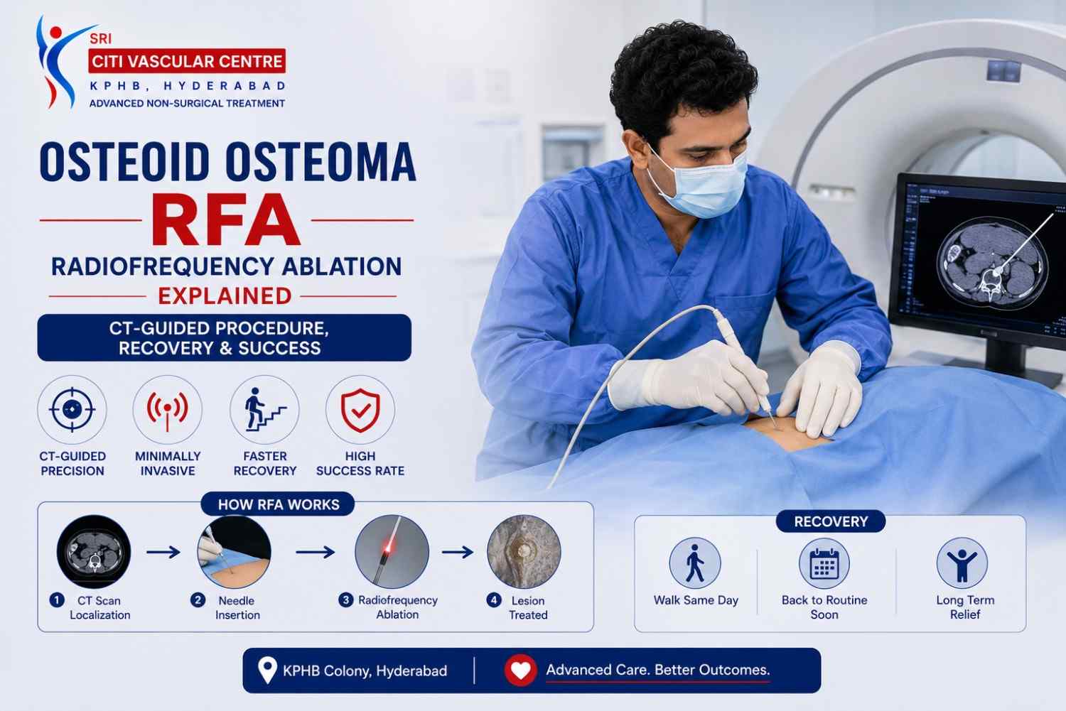
QUICK ANSWER What Is Osteoid Osteoma RFA and How Does CT-Guided Radiofrequency Ablation Work? Osteoid osteoma RFA is a minimally invasive, CT-guided procedure that uses controlled radiofrequency-generated heat to destroy the osteoid osteoma's nidus — the small central core that causes the pain. No open surgery. No large incision. Performed under anaesthesia in a single session. Systematic review of 3,023 patients shows high clinical success with an overall complication rate of approximately 3%. Dr. Garge FRCR (UK) | Citi Vascular Centre, KPHB, Hyderabad. Call +91-73375 83901. |
If you or your child has been diagnosed with an osteoid osteoma — a small, painful benign bone lesion that causes that characteristically severe night pain — and you have been told that it needs treatment, radiofrequency ablation (RFA) is very likely the first treatment your specialist will discuss. For most appropriately selected osteoid osteomas, CT-guided RFA is the evidence-based first-line interventional treatment — more precise, less invasive, and faster-recovering than open surgical removal of the affected bone.
This page explains what osteoid osteoma RFA is, how CT guidance makes the procedure accurate and safe, the five steps of the procedure, what recovery looks like, the evidence on success rates and risks, and how RFA compares with the newer microwave ablation technique. At Citi Vascular Centre, KPHB Colony, Hyderabad, Dr. Shaileshkumar Garge provides CT-guided ablation for osteoid osteoma as part of a dedicated bone-lesion interventional programme.
Osteoid Osteoma RFA Consultation — Citi Vascular Centre, KPHB, Hyderabad
Call +91-73375 83901 | WhatsApp 73375 83901 | citivascularcentre.com | Mon–Sat 9AM–6PM
|
Feature |
Detail |
|
What It Treats |
Osteoid osteoma — benign bone-forming lesion with a central nidus |
|
Procedure |
CT-guided percutaneous radiofrequency ablation of the nidus |
|
Incision |
Percutaneous — tiny needle access, no open surgical incision |
|
Anaesthesia |
Spinal or General anaesthesia, or conscious sedation — depends on age, lesion location, and planned approach |
|
Guidance |
CT imaging throughout — confirms probe tip position within the nidus before energy delivery |
|
Primary Success Rate |
96.1% (long-term study) | 91.7% after first session (systematic review of 3,023 patients) |
|
Complication Rate |
Approximately 3% overall (systematic review) — skin burns the most frequently reported |
|
Pain Relief |
97% of patients achieve significant relief | Often pain-free within days of successful ablation |
|
Recovery |
Faster than open surgery — timeline depends on lesion location and weight-bearing status |
|
Repeat RFA |
Safe and effective if residual or recurrent osteoid osteoma detected on follow-up imaging |
An osteoid osteoma is a benign (non-cancerous) bone-forming lesion — small in size but disproportionately painful. It is most commonly seen in children, teenagers, and young adults, with the femur (thigh bone), tibia (shin bone), and spine among the most frequently affected sites. Despite its small size — typically less than 1.5cm — it generates significant localised pain that classically worsens at night and is often dramatically relieved by aspirin or NSAIDs.
At the centre of the lesion is the nidus — a small, distinct area of immature bone and fibrovascular tissue that is responsible for the pain through prostaglandin production and nerve stimulation. The nidus is the target of all osteoid osteoma treatment: whether by RFA, microwave ablation, or surgical excision. CT imaging is the gold standard for identifying the nidus — it appears as a small lucent (dark) central area, often surrounded by a zone of reactive bone sclerosis (thickening). Accurate identification of the nidus is essential before any treatment is planned.
Radiofrequency ablation (RFA) is a thermal ablation technique that delivers controlled radiofrequency electrical energy through a specialised probe to the target tissue. The alternating current generates frictional heat in the tissue immediately surrounding the probe tip — reaching temperatures sufficient to cause irreversible thermal damage to the target cells. In the context of osteoid osteoma, the target is the nidus: the probe tip is positioned within the nidus, and controlled heat is applied to destroy the pain-generating nidus tissue while minimising damage to the surrounding healthy bone.
CT guidance is not optional in osteoid osteoma RFA — it is essential. The nidus is often very small (3–15mm) and may lie within a cortical bone tunnel that is difficult to visualise except with cross-sectional imaging. CT allows the operator to: confirm the exact position of the nidus before the procedure; plan the precise needle track to reach the nidus while avoiding critical adjacent structures (nerves, vessels, joints); and verify the probe tip position within the nidus before delivering radiofrequency energy. Without CT confirmation of probe position, the risk of treating adjacent normal bone or missing the nidus entirely is substantially higher.
Why is the nidus important? The nidus is the pain source — it produces prostaglandins and contains nerve fibres that generate the characteristic osteoid osteoma pain. Completely destroying the nidus by thermal ablation eliminates the pain source. An incomplete ablation that misses part of the nidus produces incomplete pain relief or early recurrence — which is why CT-confirmed probe positioning within the nidus before energy delivery is the critical quality step in osteoid osteoma RFA.
|
1 |
CT Planning and Nidus Localisation A pre-procedure CT scan precisely locates the nidus — confirming its position within the bone, measuring its depth from the skin surface, identifying adjacent nerves, joints, and blood vessels, and planning the exact needle entry point and trajectory to reach the nidus with the minimum-risk approach. |
|
2 |
Anaesthesia Appropriate anaesthesia is administered — Spinal or general anaesthesia in most paediatric patients and many adults; conscious sedation in selected cooperative adults for accessible lesions. The patient is positioned on the CT table to provide optimal access to the planned entry point. |
|
3 |
Percutaneous Probe Placement Under CT Guidance A bone access needle is advanced through the skin and through the overlying bone cortex to the nidus under stepwise CT confirmation. Position is verified by CT after each advancement — the probe tip's position within the nidus is confirmed before energy delivery begins. For cortical bone access, a drill biopsy needle is typically used to create the channel. |
|
4 |
Radiofrequency Energy Delivery The RFA probe is connected to the radiofrequency generator. Controlled energy is delivered over a planned duration (typically 4–6 minutes) at the target temperature. The operator monitors the generator's real-time temperature display to confirm adequate thermal ablation of the nidus. CT may be repeated during ablation to monitor progress in selected cases. |
|
5 |
Probe Removal and Discharge The probe and needle are removed. The small skin entry point requires only a simple dressing — no sutures in most cases. The patient is monitored in the recovery area. Most patients go home the same day or following morning. Written aftercare instructions and a follow-up appointment to assess pain resolution are provided before discharge. |
CT-guided RFA for osteoid osteoma has a strong safety record across large published series — but no procedure is completely without risk. A systematic review of 3,023 patients reported an overall complication rate of approximately 3%. Skin burns are among the most frequently reported complications, particularly when the lesion is superficial. Most complications are minor and manageable.
|
Possible Complication |
Frequency |
Notes |
|
Skin or soft-tissue thermal injury (burn) |
Most common — reported complication |
Risk higher for superficial lesions. Thermal protection techniques and careful power delivery reduce risk. |
|
Temporary post-procedure pain and swelling |
Common — expected |
The expected inflammatory response to thermal ablation — managed with analgesics. Distinct from the original osteoid osteoma pain. |
|
Infection |
Rare — < 1% |
Sterile technique and skin preparation minimise infection risk. |
|
Nerve injury |
Uncommon — varies by location |
Risk is higher when the lesion lies adjacent to a named nerve. Careful pre-procedure planning and thermal protection reduce this risk. |
|
Incomplete ablation — residual nidus |
8.3% primary failure (systematic review) |
Residual or recurrent nidus may be detectable on follow-up imaging. Repeat RFA is effective in most cases. |
|
Bone fracture at treatment site |
Rare |
More relevant for lesions in weight-bearing locations with reactive sclerosis. Weight-bearing restrictions post-procedure reduce this risk. |
RFA has a high primary success rate — but incomplete ablation of the nidus or true recurrence can occur. The systematic review of 3,023 patients reported a primary treatment-failure rate of 8.3%. The good news is that repeat RFA is effective: the secondary failure rate (after a second RFA session) drops to 3.1%, according to the same review. A long-term study reported an overall primary success rate of 96.1%, with successful retreatment in those who required a second ablation.
If pain persists or returns after RFA, it is important to have this properly assessed rather than assumed to be osteoid osteoma recurrence — because post-ablation pain from the normal inflammatory response is expected and transient, while true residual nidus requires imaging confirmation. Follow-up CT or MRI at the planned interval allows this distinction. When residual or recurrent nidus is confirmed, repeat RFA is the standard next step.
Q1: What is osteoid osteoma RFA?
Osteoid osteoma RFA is a minimally invasive, CT-guided procedure that uses controlled radiofrequency-generated heat delivered through a percutaneous probe to destroy the nidus — the small pain-causing central core of the osteoid osteoma. No open surgery, no large incision. Most patients go home the same day. Systematic review evidence across 3,023 patients demonstrates high effectiveness and a low overall complication rate. Available at Citi Vascular Centre, KPHB. Call +91-73375 83901.
Q2: Is CT-guided RFA safe for osteoid osteoma?
Yes — CT-guided RFA has an excellent safety record across large published series. A systematic review of 3,023 patients reported an overall complication rate of approximately 3%, with skin burns the most common complication. For lesions near critical structures (nerves, joints), additional precautions are used. Specialist experience and pre-procedure CT planning are the most important safety factors. RFA should not be performed without CT guidance and appropriate anatomical assessment.
Q3: Will the osteoid osteoma pain go away after RFA?
Yes — in the vast majority of successfully treated cases. One published study reports significant pain relief in 97% of patients, with many becoming pain-free within the first days after ablation. The characteristic night pain — which is often severe — typically improves rapidly once the nidus has been destroyed. Some temporary post-procedure pain from the ablation response is expected and different in character from the original osteoid osteoma pain. It settles over 1–2 weeks.
Q4: How long does recovery take after osteoid osteoma RFA?
Recovery is typically faster than open surgical excision. Most patients are home within 24 hours. Return to desk work or school: 3–7 days for most patients. Return to full physical activity and sports: depends on lesion location — for weight-bearing bones (femur, tibia), temporary activity restriction of 4–6 weeks is typically recommended. For non-weight-bearing lesions, return to full activity is usually within 2–3 weeks. Dr. Garge provides individualised post-procedure guidance specific to your lesion's location.
Q5: Can osteoid osteoma return after RFA?
Primary treatment failure (residual nidus not completely ablated) occurs in approximately 8.3% of cases (systematic review). True late recurrence is less common. When residual or recurrent osteoid osteoma is confirmed on follow-up CT, repeat RFA is safe and effective — the secondary failure rate drops to 3.1% after a second session. Persistent pain after RFA should be properly evaluated with imaging before assuming recurrence — post-ablation inflammatory pain is common and temporary.
Q6: Is RFA better than surgery for osteoid osteoma?
For most appropriately diagnosed and accessible osteoid osteomas, CT-guided percutaneous RFA is the preferred first-line treatment over open surgery — because it avoids a large incision, removes only the nidus rather than a larger bone segment, carries lower blood loss risk, and allows faster recovery. Surgery may still be considered when the diagnosis is uncertain, when the nidus cannot be safely accessed percutaneously, or when other clinical factors favour a surgical approach. The choice should follow specialist assessment.
Q7: Is RFA or microwave ablation better for osteoid osteoma?
A recent comparative study found no significant difference in clinical success between CT-guided RFA and microwave ablation for osteoid osteoma. RFA has a longer-established evidence base (3,023 patient systematic review). Microwave ablation has a growing evidence base with comparable success rates (95.8% in a 2025 systematic review). The right choice depends on lesion characteristics, anatomy, and the operator's specific expertise with each modality — not on which technique sounds newer.
Q8: Can my child have RFA for osteoid osteoma?
Yes — osteoid osteoma most commonly occurs in children, teenagers, and young adults, and RFA is well-established across all age groups. Younger children typically receive general anaesthesia rather than conscious sedation. Paediatric dosing adjustments are made for the anaesthetic agents. Parental consent is obtained. The evidence base for RFA in paediatric osteoid osteoma is strong — it avoids the scarring, bone resection, and recovery burden of open surgical excision, which matters particularly for young patients.
Q9: What happens if the first RFA session does not completely work?
If pain does not adequately resolve after RFA, or if follow-up imaging detects a residual nidus, a repeat RFA session is the standard next step. Repeat RFA is safe, technically feasible, and effective — the systematic review reports a secondary failure rate of only 3.1% after a second session. The decision to retreat is made after reviewing follow-up CT or MRI and clinical symptoms — not based on persistent post-ablation inflammatory discomfort alone, which is expected and temporary.
Q10: Where can I have CT-guided RFA for osteoid osteoma in Hyderabad?
Citi Vascular Centre, KPHB Colony, Road No. 1, Hyderabad, led by Dr. Shaileshkumar Garge — FRCR (UK), FNVIR (CMC Vellore), EBIR (Spain) — provides CT-guided thermal ablation (RFA and microwave ablation) for osteoid osteoma. Bring your recent CT scan and clinical reports for a consultation. Treatment is individualised based on nidus location and patient-specific factors. Call +91-73375 83901 or WhatsApp 73375 83901. Mon–Sat 9AM–6PM.
|
Reference |
Key Finding |
|
Tordjman M, et al. European Radiology. 2020. |
Systematic review — 3,023 patients. Primary failure 8.3%, secondary failure 3.1%. Overall complication rate ~3%. High effectiveness of CT-guided RFA for osteoid osteoma. |
|
Martel Villagrán J, et al. Skeletal Radiology. 2020. |
Systematic review of percutaneous thermal ablation for osteoid osteoma. High safety and effectiveness supporting RFA as first-line minimally invasive treatment. |
|
Abdalla BA, et al. Systematic review. 2025. |
Microwave ablation for osteoid osteoma — 143 cases. 95.8% clinical success rate. Growing evidence base comparable to RFA. |
|
Comparative CT-guided RFA vs MWA study. Recent. |
High clinical success with both techniques. No significant difference in clinical success between RFA and microwave ablation in direct comparison. |
Citi Vascular Centre, KPHB Colony, Road No. 1, Hyderabad — CT-guided osteoid osteoma ablation for patients from:
Kukatpally and KPHB — 5 min | Miyapur and Bachupally — 10 min
Hitech City, Ameerpet and Madhapur — 20 min | Gachibowli and Banjara Hills — 25 min
Secunderabad and Begumpet — 25 min | Telangana and Andhra Pradesh — outstation welcome
Osteoid osteoma RFA — CT-guided radiofrequency ablation — is a well-established, minimally invasive treatment for this small but painful benign bone lesion. Supported by a systematic review of over 3,000 patients showing 96%+ clinical success rates and an overall complication rate of approximately 3%, CT-guided RFA has replaced open surgical resection as the first-line interventional treatment for most appropriately selected osteoid osteomas. The procedure destroys the pain-causing nidus through controlled heat delivered via a percutaneously placed probe — no open incision, same-day or next-day discharge, and rapid pain relief in the majority of patients.
Where the first RFA session leaves residual disease, repeat ablation is safe and effective. Microwave ablation is an emerging alternative with comparable clinical success in recent studies — the choice between the two should be individualised rather than based on technology novelty. If you or your child has a confirmed or suspected osteoid osteoma causing significant symptoms, call +91-73375 83901 or WhatsApp 73375 83901 to discuss CT-guided ablation at Citi Vascular Centre, KPHB Colony, Hyderabad.
Osteoid Osteoma RFA — CT-Guided. Minimally Invasive. Same-Day Discharge.
96%+ Success Rate | ~3% Complication Rate | Rapid Pain Relief | No Open Surgery
Dr. Shaileshkumar Garge | FRCR (UK) | FNVIR (CMC Vellore) | EBIR (Spain) | 12+ Years | 15,000+ Procedures
Call +91-73375 83901 | WhatsApp 73375 83901 | citivascularcentre.com
Citi Vascular Centre, KPHB Colony, Hyderabad | Mon–Sat 9AM–6PM | Outstation Welcome