LAST MEDICALLY REVIEWED:
June 2026 — Dr. Shaileshkumar Garge
Citi Vascular Hospital, KPHB Colony, Road No. 1, Hyderabad, Telangana 500072
QUICK ANSWER
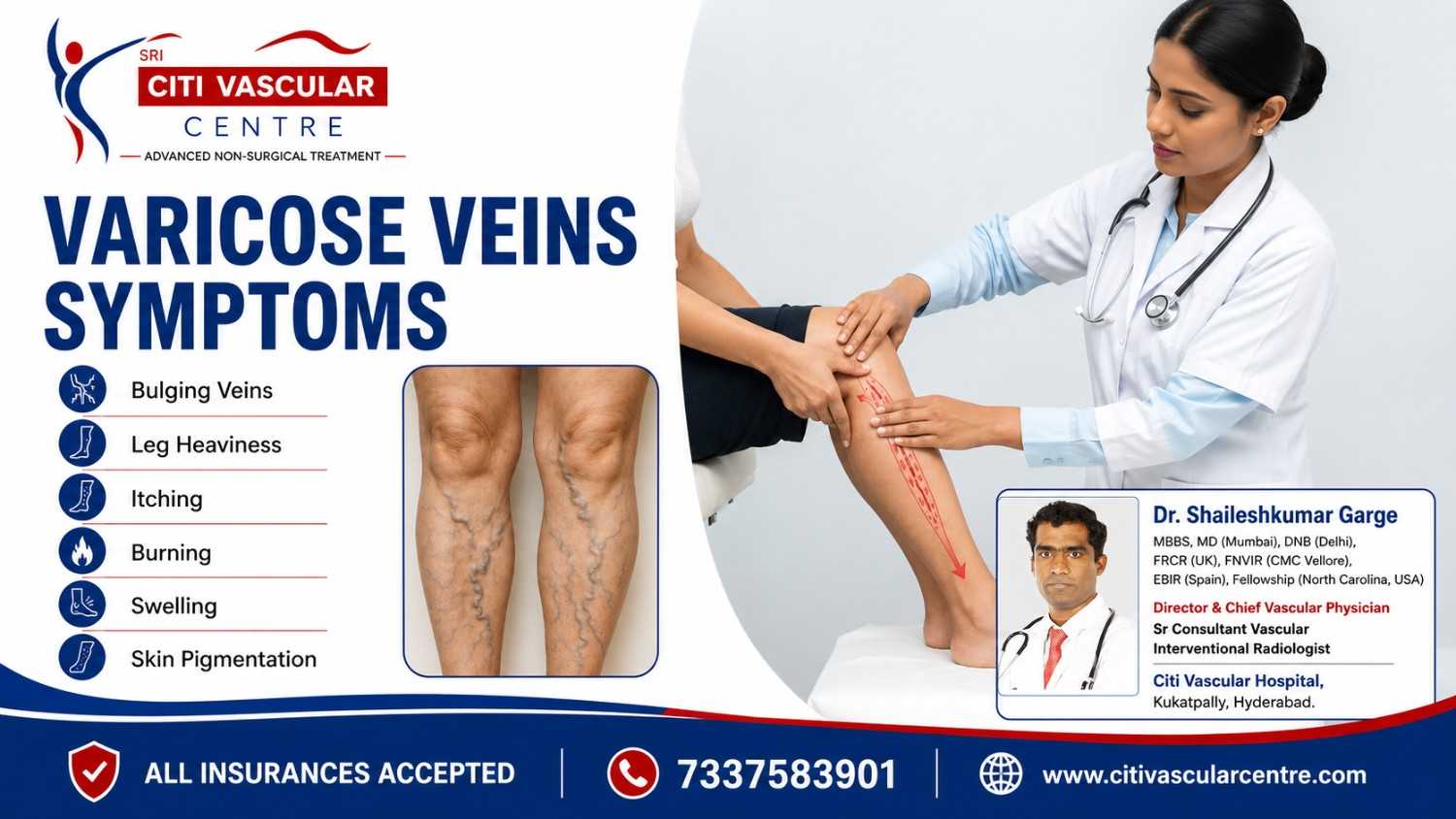
What Causes Varicose Veins and What Are the Symptoms?
Varicose veins are caused by incompetent venous valves — primarily genetic. Symptoms range from early leg heaviness and itching (C2) to skin pigmentation (C4) and active venous ulcers (C6). Most patients have symptoms before veins become visible. Do not wait for ulcers — early assessment prevents complications.
Varicose veins occur when the one-way valves inside the great saphenous vein (GSV) or small saphenous vein (SSV) become incompetent — allowing blood to flow backwards (venous reflux) and pool in the superficial leg veins under the force of gravity. The pooled blood distends the vein walls, causing the characteristic bulging, rope-like appearance. The increased venous pressure causes every symptom associated with varicose veins.
The most important clinical point about varicose vein symptoms is that many patients experience significant symptoms BEFORE their veins become visibly obvious. Leg heaviness that worsens through the day, mild ankle swelling that resolves overnight, nocturnal cramps, and itching around the ankle — these are all early venous insufficiency symptoms that frequently precede visible varicosities by months or years. Recognising these early signs allows treatment before complications develop.
This complete guide by Dr. Shaileshkumar Garge — FRCR (UK), FNVIR (CMC Vellore), EBIR (Spain) — at Citi Vascular Hospital, KPHB, Hyderabad, covers every aspect of varicose vein causes and symptoms: the biological mechanism, all causes with evidence, risk factors, the full spectrum of symptoms with clinical detail, specific symptom explanations (itching, burning, heaviness, swelling, pigmentation), the CEAP staging system, and how disease progresses if untreated.
Concerned About Varicose Vein Symptoms? Book a Duplex Doppler Evaluation
Call +91-73375 83901 | WhatsApp | citivascularcentre.com | Dr. Garge FRCR (UK) | Citi Vascular KPHB | Mon-Sat 9AM-6PM
|
Feature |
Clinical Detail |
|
Primary cause |
Incompetent venous valves in the great saphenous vein (GSV) or small saphenous vein (SSV) |
|
Most common location |
Inner thigh, calf, behind knee — anywhere the great or small saphenous vein courses |
|
First symptom in most patients |
Leg heaviness and aching worsening through the day — often before visible veins appear |
|
Strongest risk factor |
Family history (genetics) — 50-70% heritability |
|
CEAP stages |
C1 (spider veins) through C6 (active venous ulcer) — 6 clinical stages of venous disease progression |
|
Symptoms worsening pattern |
Worse after prolonged standing or sitting | Worse by evening | Improved by walking and elevation |
|
Unique symptom: itching |
Venous eczema and stasis dermatitis — most common around medial malleolus (inner ankle) |
|
Skin changes indicate |
CEAP C4 — advanced venous disease; treatment urgency increases at this stage |
|
Venous ulcer location |
Inner ankle (gaiter zone) — hallmark of CEAP C6; requires urgent specialist assessment |
|
Prevalence |
15-30% of adults | 40% of men and 32% of women (Edinburgh Vein Study) |
|
Diagnosis standard |
Duplex Doppler ultrasound — confirms reflux, grades severity, maps anatomy for treatment planning |
Normal Leg Vein Anatomy
The leg venous system has three components: (1) Deep veins — the primary drainage pathway for 90% of venous blood return, running inside the leg muscles (femoral vein, popliteal vein), (2) Superficial veins — the visible drainage network running under the skin, dominated by the great saphenous vein (GSV — inner calf and thigh) and small saphenous vein (SSV — back of calf), and (3) Perforator veins — connecting the superficial and deep systems through the calf fascia.
Varicose veins develop in the SUPERFICIAL venous system — specifically the GSV and SSV and their tributaries. These veins contain one-way bicuspid valves every 5-10cm along their length — designed to ensure blood flows upward toward the heart against gravity and prevent backflow during standing.
|
Component |
Normal Function |
In Varicose Veins |
|
Great Saphenous Vein (GSV) |
Drains inner leg/thigh superficially — 1-way valves prevent reflux |
Valve incompetence causes reflux from saphenofemoral junction down inner leg — most common source |
|
Small Saphenous Vein (SSV) |
Drains back of calf — joins popliteal vein behind knee |
SSV reflux causes posterior calf varicosities — often overlooked, assessed by Doppler |
|
Saphenous Valves |
Prevent backward blood flow during standing and Valsalva manoeuvre |
Valve cusps fail to close fully — allows blood column to fall back against gravity |
|
Tributary Veins |
Branch from GSV/SSV — drain skin and subcutaneous tissues |
Fill with pooled blood from incompetent GSV/SSV — become visibly dilated varicosities |
|
Perforators |
Transfer blood from superficial to deep system in one direction only |
Incompetent perforators allow high-pressure deep system blood to reflux into superficial veins |
Venous valve failure in the GSV and SSV is the central event in varicose vein development. The valves consist of two thin leaf-like cusps that close when blood attempts to flow backwards — preventing reflux. When these cusps fail to coapt (meet properly in the midline), a gap opens through which blood flows backwards under the force of gravity.
|
Stage |
Phase |
What Happens Biologically |
Clinical Result |
|
1 |
Valve cusp weakening |
Connective tissue in valve leaflets loses elasticity — genetic or acquired. Cusps elongate and fail to close tightly. |
Subclinical reflux begins — Doppler detectable before symptoms |
|
2 |
Retrograde blood flow |
Blood column falls back from the saphenofemoral junction toward the foot during standing — increasing hydrostatic pressure in GSV |
Venous hypertension in superficial system begins |
|
3 |
Vein wall distension |
Elevated pressure distends the vein wall — smooth muscle in the wall undergoes hypertrophy then fibrosis. Wall loses elasticity. |
Vein diameter enlarges progressively — becomes visible |
|
4 |
Valve prolapse cascade |
Once the proximal valve fails, the hydrostatic load on the next valve below increases — causing sequential failure down the vein |
Reflux extends progressively further down the leg |
|
5 |
Tributary involvement |
Increased pressure transmitted to tributary branch veins — causing their progressive dilation |
Visible varicose tributaries in thigh and calf |
|
6 |
Skin and tissue damage |
Sustained venous hypertension drives fluid and red blood cells into the interstitium — causing oedema, haemosiderin deposition, and eventually skin breakdown |
Oedema → pigmentation → lipodermatosclerosis → ulcer |
|
Cause |
Mechanism |
Clinical Evidence and Data |
|
Genetics / Family History |
Inherited connective tissue and venous wall characteristics determine valve cusp strength and vein wall elasticity |
50-70% of patients have a first-degree relative with varicose veins. If both parents affected, lifetime risk 70-80%. |
|
Female Sex / Hormonal Factors |
Oestrogen and progesterone relax vein wall smooth muscle — reducing wall tone and increasing vein distensibility |
Women affected in 32-40% of population. Risk increases with each pregnancy. OCP users have 1.5-2x increased risk. |
|
Pregnancy |
Three mechanisms: progesterone-mediated vein dilation + increased blood volume (50% rise in pregnancy) + gravid uterus compressing pelvic veins — all simultaneously increase venous pressure |
30-50% of pregnant women develop new varicose veins. Risk increases with each pregnancy. May partially resolve post-delivery. |
|
Prolonged Standing |
Static standing without walking deactivates the calf muscle pump — blood pools under gravity in the superficial veins, increasing hydrostatic pressure |
Occupations with prolonged standing (nurses, teachers, retail, chefs) have 2-3x higher varicose vein prevalence vs sedentary occupations |
|
Obesity |
Increased intra-abdominal pressure from adipose tissue impedes venous return from the legs — increasing back-pressure in saphenous veins. Adipose tissue may also release inflammatory mediators weakening vein walls. |
BMI > 30 associated with significantly higher varicose vein severity. Each 5-unit BMI increase correlates with measurable increase in venous reflux severity. |
|
Age / Ageing |
Progressive loss of elastin and collagen in vein walls and valve cusps with age — reduces structural integrity. Smooth muscle in vein walls loses contractile tone. |
Prevalence increases from 10-15% under age 25 to 50%+ over age 65. Age accelerates progression once disease begins. |
|
Prior Deep Vein Thrombosis |
DVT damages deep vein valves and walls during the inflammatory/resolution phase — post-thrombotic syndrome causes chronic deep venous hypertension transmitted to superficial system |
Post-thrombotic syndrome develops in 30-50% of DVT patients — often presenting with worsening varicose veins, leg swelling, and skin changes months to years later |
|
Sedentary Lifestyle |
Calf muscle atrophy reduces calf pump efficiency — less effective venous return propulsion. Prolonged sitting increases popliteal venous pressure. |
Regular walking maintains calf muscle pump function. Sedentary individuals have consistently higher varicose vein symptom scores. |
|
Factor |
Relative RiskRisk |
Notes |
|
Both parents have varicose veins |
Very High (70-80% lifetime) |
Strongest single predictor of varicose vein development — genetic counselling may be appropriate |
|
One parent with varicose veins |
High (40-50% lifetime) |
Routine annual examination and Doppler assessment if symptomatic in adults with family history |
|
Female sex |
1.5-2x vs age-matched males |
Oestrogen and progesterone effects on vein wall tone. Parity (number of pregnancies) is a sub-factor. |
|
Two or more pregnancies |
High — each pregnancy increases risk |
Permanent valve damage from prolonged elevated venous pressure is cumulative with each pregnancy |
|
Obesity (BMI > 30) |
Moderate-High (1.5-2x) |
Dose-response relationship — higher BMI correlates with more severe disease |
|
Standing occupation (4+ hours daily) |
Moderate (1.5-2.5x) |
Teachers, nurses, chefs, retail, hairdressers, security personnel, healthcare workers |
|
Age > 50 years |
Moderate (progressive) |
Risk doubles each decade after age 40 — age-related valve and vein wall degeneration |
|
Prior DVT |
High for post-thrombotic syndrome |
Deep venous damage causes chronic hypertension transmitted to superficial varicosities |
|
Sedentary lifestyle (< 30 min exercise daily) |
Low-Moderate |
Calf muscle pump atrophy — reduced venous return efficiency |
|
Tall stature (height > 175cm) |
Low-Moderate |
Greater venous column height increases hydrostatic pressure in leg veins during standing |
Clinical Insight: Many patients experience significant varicose vein symptoms for months or even years BEFORE any visible veins appear. This is because venous hypertension causes symptoms at the tissue level before vein diameter increases enough to be visible through the skin. These pre-visible symptoms are an important early warning sign that should not be attributed to 'tiredness' or 'aging'.
|
Early Symptom |
When It Occurs |
Why It Happens at This Stage |
|
Leg heaviness and fatigue by end of day |
Often first symptom — before visible veins |
Blood pooling in GSV increases weight-bearing venous pressure — leg muscles feel 'loaded' by the end of the day. Improves overnight. |
|
Mild ankle swelling that resolves overnight |
Early stage — may precede visible veins |
Venous hypertension drives fluid from capillaries into interstitium during the day. Overnight horizontal position allows reabsorption. |
|
Nocturnal leg cramps |
Often before visible veins appear |
Venous congestion during day causes local electrolyte disturbances and muscle irritability at night. Most common in calf muscles. |
|
Restless legs — urge to move at night |
May precede visible varicosities |
Venous hypertension and poor limb perfusion creates uncomfortable sensations requiring movement for relief. Associated with delayed sleep onset. |
|
Itching around inner ankle |
Can precede skin changes by months |
Subclinical stasis dermatitis from venous hypertension causes pruritus before visible eczema or skin changes develop |
|
Mild aching behind the knee or inner calf |
Early reflux symptom |
SSV reflux causes localised posterior calf aching. GSV reflux causes medial (inner) calf aching below the knee. |
|
Symptom |
CEAP Stage |
Urgency |
Clinical Notes |
|
Visible bulging veins |
C2 |
Evaluate |
Blue-purple rope-like veins visible through skin. Worse standing. GSV distribution: inner calf and thigh. SSV: posterior calf. |
|
Leg heaviness and aching |
C2-C3 |
Evaluate |
Classic symptom. Worsens through the day, after prolonged standing, and by evening. Relieves with walking and leg elevation. |
|
Ankle and lower leg swelling |
C3 |
Evaluate |
Pitting oedema — typically worse by evening, resolved by morning. Persistent swelling suggests C3+ disease needing prompt assessment. |
|
Night cramps (nocturnal) |
C2-C3 |
Evaluate |
Calf muscle cramps waking patient at night — caused by venous congestion and muscle irritability from chronic venous hypertension. |
|
Itching (venous eczema) |
C2-C4 |
Evaluate soon |
Pruritus around ankle and lower leg — caused by stasis dermatitis. Scratching damages fragile skin and risks ulcer formation. |
|
Burning sensation along veins |
C2-C3 |
Evaluate |
Warm, burning feeling along the course of superficial varicosities — caused by venous inflammation and stasis |
|
Skin pigmentation (brown discolouration) |
C4a |
Urgent — evaluate within weeks |
Haemosiderin deposition in skin from red blood cell extravasation. Permanent once established. Indicates significant venous hypertension. |
|
Venous eczema (dermatitis) |
C4a |
Urgent |
Weeping, crusting, inflamed skin around ankle — acute inflammation from venous hypertension. High risk of skin breakdown. |
|
Lipodermatosclerosis |
C4b |
Urgent |
Hard, indurated, woody skin around lower leg — fibrotic scarring of subcutaneous fat from chronic venous congestion. Pre-ulcer state. |
|
Atrophie blanche |
C4c |
Urgent |
White, shiny, scar-like skin patches surrounded by dilated capillaries — hallmark of severe chronic venous hypertension. High ulcer risk. |
|
Active venous ulcer |
C6 |
URGENT |
Non-healing wound on inner ankle — typically painless with moist base, irregular edges, surrounded by pigmented skin. Requires urgent specialist care. |
|
Spontaneous varicosity bleeding |
Any stage |
EMERGENCY |
Rupture of large varicosity — lie down, elevate leg, apply firm pressure. Go to emergency if uncontrolled. |
The Mechanism of Venous Itch
Itching (pruritus) associated with varicose veins is one of the most frequently misunderstood symptoms — often dismissed as dry skin or insect bites. It is actually a specific clinical entity called stasis dermatitis (venous eczema) caused directly by venous hypertension in the skin.
|
Stage |
What Happens in the Skin |
Clinical Appearance |
|
Venous Hypertension |
Sustained high pressure in skin capillaries forces fluid and proteins into the surrounding tissues |
Skin around ankle feels heavy, warm, and starts to itch |
|
Inflammatory Response |
White blood cells infiltrate the skin in response to protein extravasation — releasing inflammatory mediators including histamine |
Intense itching develops — predominantly around the medial malleolus (inner ankle) |
|
Stasis Dermatitis Develops |
Chronic inflammation damages the epidermal barrier — skin becomes dry, scaly, and weeping in some areas |
Visible venous eczema — red, scaly, crusting skin around the ankle (CEAP C4a) |
|
Haemosiderin Deposition |
Red blood cells leak through damaged capillaries into the skin — haemoglobin breaks down to haemosiderin |
Brown pigmentation develops beneath the areas of itch (permanent once established) |
|
Skin Breakdown Risk |
Scratching the fragile, eczematous skin creates microtrauma — a portal for venous ulcer formation |
Do NOT scratch. Apply emollient and seek specialist assessment urgently. |
Important: Varicose vein itching around the ankle should NEVER be treated with topical steroids long-term without specialist assessment. Steroid overuse causes skin thinning — increasing the risk of the very skin breakdown (ulceration) that is being tried to prevent. If you have persistent ankle itching with visible varicose veins, book a Doppler assessment at Citi Vascular Hospital, KPHB.
A burning, warm, or tingling sensation along the course of varicose veins is another specific symptom caused by venous inflammation (phlebitis-like reaction within distended veins) and the nerve irritation that occurs from sustained venous congestion in the surrounding tissues.
|
Type of Burning Sensation |
What It Means Clinically |
|
Burning along vein course — gradual onset |
Venous inflammation (phlebitis) inside the varicosity — chronic distension and stasis creates local inflammatory reaction along the vein wall |
|
Burning sensation at ankle and lower leg |
Stasis dermatitis early stage — same mechanism as itch but the patient perceives it as burning before the itching stage develops |
|
Sudden onset burning and redness along a vein |
Superficial thrombophlebitis — clot forming inside a varicosity causes acute inflammatory reaction. Seek urgent assessment: call +91-73375 83901. |
|
Burning that worsens in heat (hot weather, hot bath) |
Heat dilates veins further — increasing pooling and venous pressure temporarily. Patients often find symptoms significantly worse in summer and after hot showers. |
|
Burning at night |
Venous congestion accumulated during the day causes tissue irritation peaking in the evening. Leg elevation provides relief. |
Why Do Legs Feel Heavy with Varicose Veins?
Leg heaviness is the most common varicose vein symptom — reported by over 80% of symptomatic patients. It is caused by blood pooling in the distended superficial veins under gravity. The weight of the pooled venous blood, combined with the inflammatory fluid forced into the interstitium by elevated venous pressure, creates a genuine physical weight sensation in the lower leg.
|
Heaviness Pattern |
Clinical Significance |
|
Worsens through the day |
Blood accumulates in dilated leg veins progressively during standing or sitting — most pronounced by late afternoon and evening |
|
Relieves with walking |
Walking activates the calf muscle pump — propels pooled blood upward and reduces venous pressure. Even 10 minutes provides noticeable relief. |
|
Relieves with leg elevation |
Gravity assists venous drainage from superficial leg veins when legs are elevated above heart level. Most patients sleep better with legs slightly elevated. |
|
Relieves overnight |
Horizontal sleeping position eliminates hydrostatic pressure. Blood drains passively during sleep. Morning is the best time of day for most patients. |
|
Does NOT relieve with rest (sitting) |
Sitting without leg elevation or movement does not reduce venous pooling — it may worsen it if the popliteal vein is compressed at the knee by the chair edge. |
Swelling from Varicose Veins — Mechanism and Pattern
Venous oedema (swelling from varicose veins) develops when the hydrostatic pressure in the capillaries exceeds the oncotic pressure that keeps fluid inside the vessel — forcing protein-rich fluid into the interstitium (tissue spaces). This is pitting oedema — pressing on the swollen area leaves an indentation.
|
Swelling Feature |
Varicose Vein Oedema |
Other Causes to Distinguish |
|
Location |
Ankle and lower leg — typically below the knee |
Heart failure: bilateral, extends to thighs | Lymphoedema: non-pitting, may extend to toes |
|
Daily pattern |
Worsens through the day — absent or minimal in the morning |
Heart failure: present on waking | Lymphoedema: persistent without daily variation |
|
Response to elevation |
Resolves significantly within 1-2 hours of leg elevation |
Lymphoedema does not fully resolve with elevation | Heart failure partially improves |
|
Pitting |
Yes — finger pressure leaves indentation that persists 30-60 seconds |
Lymphoedema: non-pitting (fibrotic) | DVT: firm, non-pitting unilateral swelling |
|
Associated features |
Visible varicose veins, aching, heaviness, and skin changes |
Confirm with duplex Doppler at Citi Vascular KPHB — distinguishes venous from non-venous causes |
Skin changes associated with varicose veins represent CEAP C4 disease — advanced chronic venous insufficiency where sustained venous hypertension has begun to cause irreversible changes to the skin and subcutaneous tissues of the lower leg. These are clinically significant warning signs that the disease is progressing toward ulceration if untreated.
|
Skin Change |
CEAP Sub-Stage |
Mechanism |
Clinical Significance |
|
Brown/tan pigmentation around inner ankle |
C4a |
Haemosiderin deposition — iron from extravasated red blood cells deposits in dermis |
Permanent once established. Indicates years of venous hypertension. |
|
Venous eczema (stasis dermatitis) |
C4a |
Inflammatory response to protein extravasation — epidermal barrier disruption |
Weeping, crusting, intensely itchy skin. High risk of infection. Treat underlying venous disease urgently. |
|
Lipodermatosclerosis |
C4b |
Fibrosis of subcutaneous fat from chronic venous inflammation — tissue becomes hard, indurated, and bound down |
Skin feels like wood. Leg adopts inverted champagne bottle appearance. Pre-ulcer state. |
|
Atrophie blanche |
C4c |
Avascular white plaques from capillary thrombosis — surrounded by dilated capillaries (telangiectasias) |
Highly predictive of imminent ulceration. Urgent specialist assessment required. |
|
Corona phlebectatica |
C4c |
Fan-shaped telangiectasias (spider veins) spreading across the inner ankle and foot |
Marker of advanced perimalleolar venous hypertension. Significant risk of progression. |
Treatment urgency: Any patient with CEAP C4 skin changes should not wait. Skin changes indicate the disease has progressed beyond simple varicose veins to chronic venous insufficiency with significant tissue damage. Without treating the underlying reflux, progression to active venous ulceration (C6) becomes a significant risk. Book an urgent assessment at Citi Vascular Hospital, KPHB: +91-73375 83901.
A venous leg ulcer is the end-stage complication of untreated chronic venous insufficiency — representing CEAP C6 disease. It develops when sustained venous hypertension finally overwhelms the skin's reparative capacity — typically in the gaiter zone (inner ankle) where the skin is most exposed to perforator-driven venous hypertension. Venous ulcers are slow to heal, painful, and have high recurrence rates without treating the underlying venous reflux.
|
Feature of Venous Ulcer |
Clinical Details |
|
Location |
Inner ankle (medial malleolus) — gaiter zone. This is the hallmark location distinguishing venous from arterial ulcers. |
|
Wound appearance |
Shallow wound with moist, red or yellow wound bed. Irregular, sloping edges. Surrounding skin with heavy brown pigmentation and lipodermatosclerosis. |
|
Pain level |
Usually mild-moderate — much less painful than arterial ulcers. Some patients have minimal pain — which can falsely reassure them the wound is not serious. |
|
Size and chronicity |
Can range from 1cm to large wounds covering the entire gaiter zone. Average healing time without reflux treatment: 6-12 months. |
|
Healing with compression alone |
Possible but slow and high recurrence — ESCHAR trial showed 50-60% 1-year recurrence with compression alone without treating reflux |
|
Healing after varicose vein treatment |
Significantly faster healing with endovenous reflux treatment (RFA/EVLT/VenaSeal) combined with wound care. ESCHAR trial: treating reflux halved ulcer recurrence rate at 4 years. |
For detailed information on varicose vein treatment options including RFA, EVLT (laser), VenaSeal (glue), and foam sclerotherapy:-> Treatment comparison: citivascularcentre.com/surgery-vs-evlt-vs-venaseal-for-varicose-veins-> RFA in detail: citivascularcentre.com/rfa-treatment-for-varicose-veins-hyderabad-> VenaSeal in detail: citivascularcentre.com/glue-treatment-for-varicose-veins-hyderabad
|
CEAP |
Stage Name |
Clinical Description |
Common Patient Complaint |
Treatment Urgency |
|
C0 |
No visible disease |
No signs on examination but may have symptoms from deep venous disease |
Unexplained leg aching |
Investigate if symptomatic |
|
C1 |
Telangiectasias (spider veins) |
Dilated intradermal venules < 1mm — red/blue thread veins visible at skin surface |
Cosmetic concern | mild itch |
Elective — sclerotherapy available |
|
C2 |
Varicose Veins |
Dilated subcutaneous veins > 3mm — visible blue-green bulging vein clusters in thigh and calf |
Heaviness, aching, visible veins, night cramps |
Evaluate — treat when symptomatic |
|
C3 |
Oedema |
Pitting ankle oedema from venous hypertension — worsens through day, resolves overnight |
Ankle swelling by evening | tight shoes by end of day |
Evaluate and discuss treatment |
|
C4a |
Skin Changes (mild) |
Haemosiderin pigmentation (brown skin) and/or venous eczema around the ankle |
Brown ankle skin | itching and dermatitis |
Prompt treatment recommended |
|
C4b |
Skin Changes (severe) |
Lipodermatosclerosis (hard, fibrotic skin) and/or atrophie blanche (white scarring) |
Hard, woody skin | white patches | ankle tenderness |
Urgent — high ulcer risk |
|
C5 |
Healed Ulcer |
Previously open venous ulcer that has healed but skin remains abnormal |
History of previous leg wound | chronic skin damage |
Urgent — high recurrence risk |
|
C6 |
Active Venous Ulcer |
Open non-healing wound in the gaiter zone — active venous ulceration |
Non-healing wound on inner ankle |
Emergency assessment required |
Clinical Note: The CEAP classification is not just academic — it directly determines treatment urgency and approach. CEAP C2-C3 patients are offered treatment on a selective basis. CEAP C4-C6 patients are recommended treatment as a medical priority to prevent further progression. Dr. Garge assesses every patient's CEAP stage at duplex Doppler consultation at Citi Vascular Hospital, KPHB.
|
1 |
C1: Spider Veins Appear Incompetent valve begins — increased pressure transmitted to smallest capillaries and venules. Fine red or blue thread veins appear at skin surface. Minimal symptoms. Often cosmetic concern only at this stage. |
|
2 |
C2: Varicose Veins Become Visible GSV/SSV reflux established — tributary veins fill and dilate to > 3mm. Blue-green rope-like veins visible through skin. Leg heaviness, aching, and nocturnal cramps begin. |
|
3 |
C3: Ankle Oedema Venous hypertension begins forcing fluid from capillaries into tissue. Pitting ankle oedema develops — worst by evening, resolves overnight. Shoes tighter in the afternoon. Swelling may extend to lower calf. |
|
4 |
C4a: Skin Pigmentation and Eczema Red blood cells leak into skin — haemosiderin deposits create permanent brown pigmentation around ankle. Venous eczema develops — itching, scaling, and inflammation. Skin barrier begins to break down. |
|
5 |
C4b: Lipodermatosclerosis and Atrophie Blanche Chronic inflammation causes fibrotic hardening of the subcutaneous fat (lipodermatosclerosis) — skin becomes hard and board-like. White, avascular plaques appear (atrophie blanche). Ulcer risk now HIGH. |
|
6 |
C5-C6: Venous Ulcer Formation Skin integrity finally fails under sustained venous pressure — typically triggered by minor trauma to fragile skin. An open non-healing wound (venous ulcer) develops on the inner ankle. Can take months to years to heal without treating the underlying reflux. |
Not every patient progresses through all stages — and progression is NOT inevitable. Early treatment at C2-C3 prevents the entire C4-C6 progression. The goal of treatment is not just symptom relief but PREVENTION of skin damage and ulceration. The ESCHAR trial demonstrated that treating underlying venous reflux at any stage significantly reduces progression risk.
High-Risk Occupations
|
Occupation |
Risk Level |
Reason |
|
Teachers / Lecturers |
Very High |
Standing 6-8 hours daily with limited walking opportunity — maximum static venous pressure |
|
Nurses / Healthcare workers |
Very High |
Prolonged standing plus shift patterns without adequate rest — high cumulative venous pressure load |
|
Retail and shop workers |
High |
Prolonged standing on hard floors — particularly hard concrete or tiles without anti-fatigue matting |
|
Chefs and kitchen staff |
High |
Standing 8-12 hours in hot environments — heat dilates veins further, worsening pooling on an already-standing-stressed system |
|
IT professionals and office workers |
Moderate-High |
Prolonged sitting deactivates calf pump + popliteal vein compression from chair edge increases backpressure |
|
Hairdressers and beauticians |
High |
Prolonged standing on hard floors | poorly fitting shoes on hard surfaces |
|
Security personnel |
High |
Extended static standing shifts — often 8-12 hours with minimal walking |
|
Surgeons and dentists |
Moderate |
Extended standing during procedures — though operating on a rubber anti-fatigue mat partially mitigates risk |
Other High-Risk Groups
|
Group |
Specific Risk Factors |
|
Pregnant women |
Three simultaneous risk factors: progesterone-mediated vein dilation + 50% blood volume increase + uterine pelvic vein compression. Risk increases with each pregnancy. |
|
Obese individuals (BMI > 30) |
Intra-abdominal pressure elevation + inflammatory mediators from adipose tissue + reduced exercise tolerance — all worsen venous insufficiency |
|
Prior DVT patients |
Post-thrombotic syndrome in 30-50% — deep vein valve damage causes chronic venous hypertension transmitted to superficial system |
|
Patients with strong family history |
Both parents affected = 70-80% lifetime risk. Even one parent doubles the risk vs those without family history. |
|
Symptom or Finding |
Urgency |
Action Required |
|
Active bleeding from varicose vein |
EMERGENCY |
Lie down, elevate leg, apply firm pressure. Go to ER or call emergency services if bleeding does not stop. |
|
Active venous ulcer (non-healing ankle wound) |
Urgent — within days |
Book emergency assessment at Citi Vascular Hospital, KPHB: +91-73375 83901 |
|
Sudden hardness, redness, pain along a vein |
Urgent — 24-48 hours |
Superficial thrombophlebitis — needs Doppler to rule out DVT extension |
|
Ankle skin becoming dark brown or hard |
Within 1-2 weeks |
CEAP C4 disease — pre-ulcer state. Early treatment prevents ulcer formation. |
|
Persistent swelling not improving overnight |
Within 2 weeks |
Assess for C3+ disease. Doppler confirms venous cause and guides treatment. |
|
Itching around ankles with visible varicose veins |
Within 2-4 weeks |
Stasis dermatitis — early C4a. Do not scratch. Seek assessment and treatment. |
|
Visible bulging veins with heaviness or pain |
Elective — within weeks |
Doppler assessment and treatment discussion. C2-C3 disease. |
|
Visible bulging veins with no symptoms |
Elective — within months |
Annual Doppler monitoring. Treatment when symptoms develop or progression detected. |
|
Credential |
Detail |
|
Full Name |
Dr. Shaileshkumar Garge |
|
Qualifications |
MBBS | MD (Mumbai) | DNB (Delhi) | FRCR (UK) | FNVIR (CMC Vellore) | EBIR (Spain/Europe) | Fellowship (North Carolina, USA) |
|
Role |
Director and Chief Vascular Physician | Senior Consultant Vascular and Interventional Radiologist |
|
Hospital |
Citi Vascular Hospital, KPHB Colony, Road No. 1, Hyderabad, Telangana 500072 |
|
Varicose Vein Expertise |
Diagnosis | CEAP staging | Duplex Doppler assessment | RFA | EVLT | VenaSeal | MOCA | Foam Sclerotherapy | Micro-Phlebectomy |
|
Experience |
12+ years | 15,000+ minimally invasive vascular procedures across Hyderabad and Telangana |
Q1: What is usually the first sign of varicose veins?
The most common first sign of varicose veins is leg heaviness or aching that worsens through the day and improves overnight or with walking — typically appearing months or years BEFORE visible veins develop. Mild ankle swelling, nocturnal cramps, and itching around the inner ankle are also early indicators. These pre-visible symptoms are caused by venous hypertension before vein diameter increases enough to be seen.
Q2: Can varicose veins cause leg pain without visible bulging veins?
Yes — varicose vein pain frequently precedes visible veins by months or years. Venous hypertension from early GSV reflux causes leg heaviness, aching, and cramps long before veins become visible. A Doppler ultrasound can confirm reflux even when no visible varicosities are present. If you have persistent unexplained leg aching that worsens through the day, a Doppler assessment at Citi Vascular, KPHB, Hyderabad is recommended.
Q3: What does varicose vein itching feel like and where does it occur?
Varicose vein itching (stasis dermatitis or venous eczema) is most intense around the inner ankle (medial malleolus) — not along the visible varicose veins themselves. It feels like a persistent, deep itch that is difficult to relieve without scratching. The skin may appear dry, scaly, or red. It is caused by venous inflammation from sustained venous hypertension in the skin capillaries — not dry skin or an allergy.
Q4: Why do varicose vein symptoms worsen at the end of the day?
Varicose vein symptoms worsen by evening because venous blood accumulates in the superficial leg veins under gravity throughout the day. Each hour of standing or sitting without walking increases the volume of pooled blood, progressively raising venous pressure. By evening, the leg is at its most congested — maximum heaviness, swelling, aching, and burning. Overnight horizontal rest allows venous drainage, explaining why mornings are most comfortable.
Q5: What is lipodermatosclerosis and is it serious?
Lipodermatosclerosis (LDS) is the hardening and fibrosis of the subcutaneous fat in the lower leg from chronic venous inflammation — CEAP C4b disease. The skin becomes hard, wooden, and indurated — the leg acquires an inverted champagne bottle appearance. LDS indicates severe chronic venous insufficiency and is a pre-ulcer state. Without treating the underlying venous reflux, ulcer formation becomes a significant and imminent risk.
Q6: Can varicose veins cause night cramps?
Yes — nocturnal leg cramps (muscle spasms waking patients at night) are a well-recognised varicose vein symptom. Venous congestion accumulated during the day creates local electrolyte imbalances and nerve irritability in the calf muscles. The cramps typically occur in the calf and are most frequent on nights following prolonged standing or sitting. They usually reduce significantly after varicose vein treatment, though this is a secondary symptom rather than a primary diagnostic criterion.
Q7: Are varicose veins hereditary — will my children develop them?
Yes — varicose veins have 50-70% heritability. If one parent has varicose veins, a child has approximately 40-50% lifetime risk. If both parents are affected, the risk rises to 70-80%. The inherited trait involves venous wall connective tissue strength and venous valve characteristics. Children of affected parents should: maintain healthy BMI, exercise regularly, avoid prolonged static standing, and seek early assessment if symptoms develop in adulthood.
Q8: Can varicose veins appear during or after pregnancy?
Yes — varicose veins are extremely common in pregnancy, affecting 30-50% of pregnant women. Three simultaneous mechanisms drive this: progesterone relaxes vein walls, blood volume increases 50%, and the growing uterus compresses pelvic veins. Most women notice onset during the first trimester, worsening through pregnancy. Only 30-40% of pregnancy-related varicose veins fully resolve after delivery — the majority persist and worsen with subsequent pregnancies.
Citi Vascular Hospital, KPHB Colony, Road No. 1, Hyderabad — varicose vein symptom assessment, Doppler ultrasound, and CEAP staging available for patients from:
Kukatpally and KPHB — 5 min drive
Miyapur and Bachupally — 10 min
Hitech City and Madhapur — 20 min
Ameerpet and SR Nagar — 20 min
Gachibowli and Kondapur — 25 min
Secunderabad and Begumpet — 25 min
Kompally, Medchal and Alwal — 20-25 min
Telangana and AP — outstation consultations welcome
|
Hospital |
Contact |
Hours |
|
Citi Vascular Hospital |
+91-73375 83901 |
KPHB Colony, Road No. 1, Hyderabad, Telangana 500072 | Mon-Sat 9AM-6PM |
|
Online Booking |
citivascularcentre.com |
WhatsApp + online | Same-day Doppler assessment available | Insurance assisted |
SUMMARY
Varicose veins are a progressive condition caused by faulty leg vein valves, leading to backward blood flow and increased venous pressure. Symptoms may begin with leg heaviness, pain, cramps, swelling, and skin changes long before bulging veins appear. Early diagnosis is important because treatment is simpler and more effective in the initial stages, while advanced disease can lead to skin damage and venous ulcers.
At Citi Vascular Hospital, KPHB Colony, Hyderabad, Dr. Shaileshkumar Garge (FRCR UK, FNVIR, EBIR) performs a detailed Duplex Doppler evaluation to confirm venous reflux, assess disease severity, and provide personalised treatment recommendations or reassurance when intervention is not required.
Book Your Varicose Vein Assessment — Symptoms Evaluated, CEAP Staged, Doppler on Site
Citi Vascular Hospital, KPHB Colony, Road No. 1, Hyderabad
Dr. Shaileshkumar Garge | FRCR (UK) | FNVIR (CMC Vellore) | EBIR (Spain) | 12+ Years | 15,000+ Procedures
Call +91-73375 83901 | WhatsApp Now | citivascularcentre.com
Duplex Doppler | CEAP Staging | All Treatment Options | Same-Day Discharge | Insurance Assisted | Mon-Sat 9AM-6PM