LAST MEDICALLY REVIEWED:
August 2026 — Dr. Shaileshkumar Garge
Citi Vascular Hospital, KPHB Colony, Road No. 1, Hyderabad, Telangana 500072
QUICK ANSWER
How Is Venous Malformation Sclerotherapy Performed in Hyderabad?
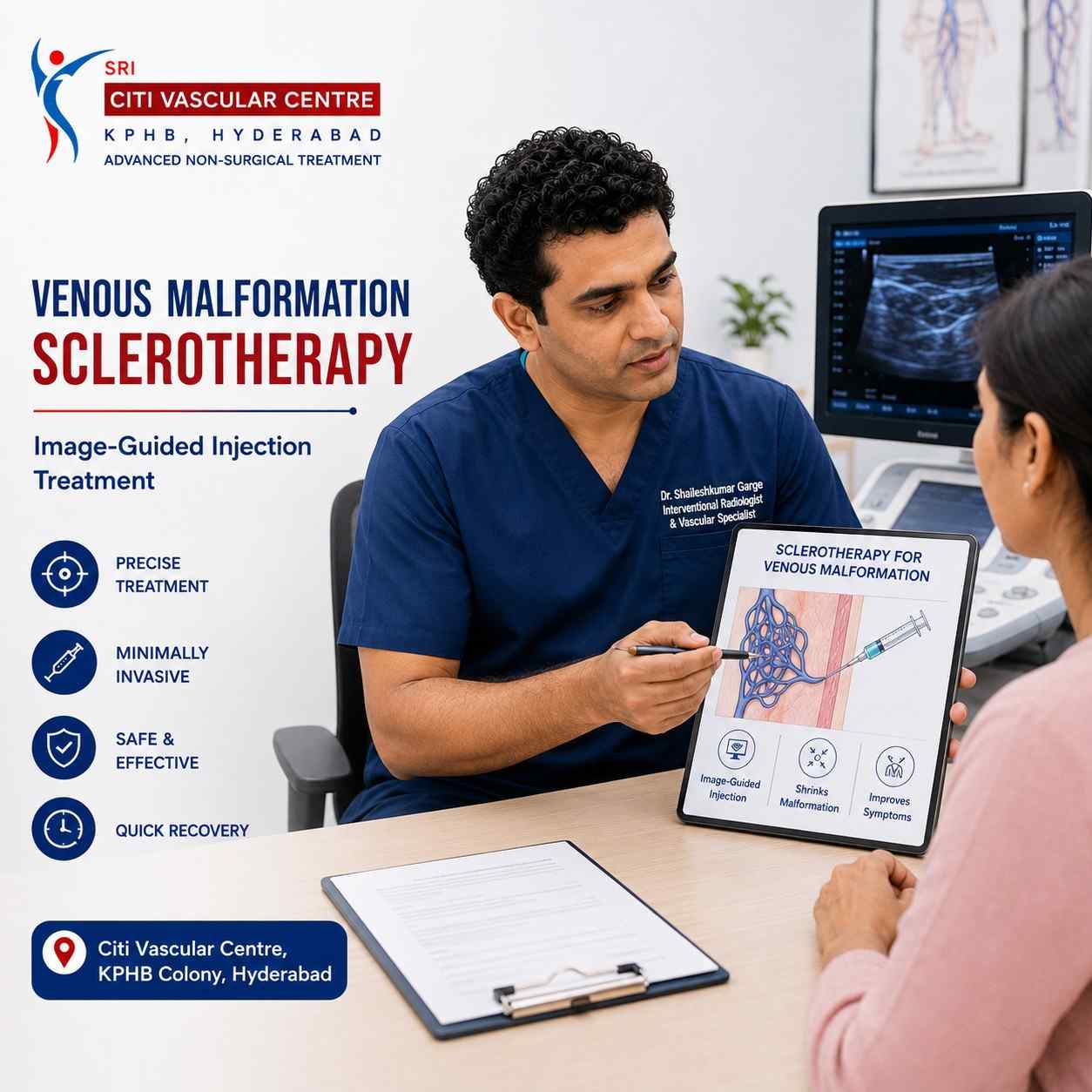
Venous malformation sclerotherapy is an image-guided procedure in which a specialised sclerosing agent is injected directly into the abnormal venous channels under real-time ultrasound (and when required, fluoroscopic) guidance. The sclerosant destroys the abnormal venous lining, causing the channels to collapse, fibrose, and gradually shrink. No major surgical incision. Most procedures are day-care. Multiple sessions are usually needed. Dr. Garge FRCR (UK) | Citi Vascular Centre, KPHB, Hyderabad. Call +91-73375 83901.
For patients with venous malformations that require active treatment — because of pain, functional limitation, cosmetic concern, or progressive enlargement — image-guided sclerotherapy is the most widely used and most evidence-supported interventional option. It avoids the need for open surgery in most cases, treats the malformation from within the abnormal venous channels themselves, preserves surrounding healthy tissue, and allows most patients to go home the same day. Yet despite being the first-line interventional treatment recommended by international vascular anomaly guidelines, sclerotherapy for venous malformation remains poorly understood by most patients who are referred for it.
The most common questions patients ask before their first sclerotherapy session are the same ones: How does it work? Will it hurt? Will one session be enough? When will I see results? What are the risks? This page answers all of those questions in full — based on the technique used at Citi Vascular Centre, KPHB, Hyderabad, and the published international evidence base for image-guided sclerotherapy. For background information on venous malformations — what they are, how they are diagnosed, and the full range of treatment options — see our Venous Malformation Treatment in Hyderabad overview page.
Venous Malformation Sclerotherapy — Citi Vascular Centre, KPHB, Hyderabad
Call +91-73375 83901 | WhatsApp 73375 83901 | citivascularcentre.com | Mon–Sat 9AM–6PM
|
Feature |
Detail |
|
Treatment Type |
Image-guided sclerotherapy — minimally invasive, no major surgical incision |
|
Used For |
Low-flow venous malformations — confirmed on Doppler USG and MRI |
|
Imaging Guidance |
Ultrasound guidance (primary) ± Fluoroscopy (for complex or deep lesions) |
|
Hospital Stay |
Day-care for most patients | Overnight or short stay for complex large VMs |
|
Anaesthesia |
Local anaesthesia + IV sedation for most | General anaesthesia for selected complex cases (children, extensive lesions, airway proximity) |
|
Procedure Duration |
30–90 minutes per session depending on VM size and complexity |
|
Sessions Required |
1 session for very small VMs | 2–5 sessions (at 4–8 week intervals) for most significant VMs | More for large/diffuse lesions |
|
Recovery |
1–3 days to light activities | 1–2 weeks before significant swelling resolves | Months for maximum shrinkage |
|
Main Goal |
Pain relief, functional improvement, volume reduction, cosmetic improvement — not necessarily complete elimination |
|
Specialist |
Interventional Radiologist — trained in image-guided vascular anomaly procedures |
Sclerotherapy for venous malformation is a minimally invasive, image-guided treatment in which a specialised chemical agent — called a sclerosant — is injected directly into the abnormal venous channels that make up the malformation. The sclerosant works by chemically irritating and damaging the inner lining (endothelium) of the abnormal venous channels from within — causing them to collapse, clot, and eventually fibrose (scar down) over the following weeks and months. As the treated channels are progressively replaced by fibrous scar tissue, the visible and palpable volume of the venous malformation reduces, and the symptoms it was causing — pain, swelling, functional limitation, cosmetic concern — improve.
The word 'sclerotherapy' comes from the Greek 'sclero' (hard) — reflecting the scarring and hardening of the treated tissue that is the mechanism of action. The key feature that distinguishes modern venous malformation sclerotherapy from older techniques is the use of real-time image guidance throughout the procedure. By inserting the needle under continuous ultrasound visualisation (and using fluoroscopic confirmation of contrast spread when appropriate), the sclerosant is delivered precisely into the target VM channels — not into adjacent normal tissue or blood vessels. This precision is what makes image-guided sclerotherapy substantially safer and more effective than older approaches involving blind injection.
Sclerotherapy is endorsed as the primary interventional treatment for most symptomatic low-flow venous malformations by the Society of Interventional Radiology (SIR), the Cardiovascular and Interventional Radiological Society of Europe (CIRSE), and the International Society for the Study of Vascular Anomalies (ISSVA). The evidence base from multiple published series confirms significant symptom improvement and lesion volume reduction in the majority of well-selected patients — while avoiding the surgical risks associated with direct excision of venous malformation tissue (particularly blood loss from the highly vascular channels).
Clinical Assessment and MRI Review
Before any sclerotherapy is planned, Dr. Garge reviews your Doppler ultrasound (confirming low-flow venous nature — no arterial signal) and your MRI (defining the three-dimensional extent of the VM, its relationship to adjacent nerves, vessels, and bone). Treatment planning is specific to the individual VM — size, location, depth, and which area is most symptomatic are all factored in.
Image-Guided Needle Placement
Under real-time ultrasound guidance, one or more needles are carefully positioned within the VM channels. The needle tip position within the abnormal venous tissue is confirmed on the ultrasound screen before any injection begins. For deeper or more complex VMs where ultrasound alone may not provide complete visualisation, fluoroscopic (X-ray) guidance is added.
Contrast Confirmation (When Appropriate)
Before the sclerosant is injected, a small amount of radiographic contrast dye is injected through the needle under fluoroscopic screening. This confirms that the needle is correctly positioned within the VM, reveals the extent of the venous channels that will be treated in this session, and importantly identifies any venous drainage pathways from the VM into larger veins — which must be accounted for in dosing.
Sclerosant Injection
The sclerosant is injected slowly and carefully under continuous imaging — the volume injected, the rate of injection, and the pressure are all controlled to maximise treatment of the target VM while minimising the risk of reflux into normal veins. Injection is paused immediately if contrast or sclerosant tracks towards a non-target vessel.
Abnormal Venous Channels Collapse
The sclerosant chemically destroys the endothelial lining of the abnormal venous channels it contacts, triggering thrombosis and inflammatory response within the treated tissue. On follow-up imaging, the treated channels appear echogenically brighter on ultrasound (indicating thrombosis) and subsequently reduce in size on serial MRI.
Compression and Monitoring
After injection, the treated area is compressed firmly for several minutes to reduce bleeding and maximise contact between the sclerosant and the channel walls. The patient is observed in the recovery area for 1–4 hours before discharge. Vital signs and the treated area are monitored throughout.
Gradual Healing and Shrinkage
Over the following 4–8 weeks, the thrombosed channels are progressively replaced by fibrous scar tissue. This is the phase during which the visible and palpable VM volume reduces. The process continues over months — which is why follow-up MRI is scheduled at 2–3 months after each session to assess response before the next session is planned.
Not every venous malformation requires sclerotherapy, and not every VM is suitable for sclerotherapy as the primary or sole treatment. The decision is made by Dr. Garge at consultation after reviewing the clinical picture, Doppler ultrasound, and MRI. The following patient profiles generally indicate appropriate candidates for sclerotherapy.
|
Suitable Profile |
Why Sclerotherapy Is Appropriate |
|
Symptomatic venous malformation — pain affecting daily life |
Pain from VM thrombosis, swelling, or pressure on adjacent structures — the most common primary indication for sclerotherapy. Published series consistently show significant pain improvement in most patients. |
|
Progressive enlargement on serial imaging |
A VM that is demonstrably growing on Doppler USG or MRI over time — treatment indicated before further enlargement causes greater functional or cosmetic impact |
|
Functional limitation — reduced range of movement, difficulty swallowing |
VMs adjacent to joints limit movement; oropharyngeal VMs affect swallowing or speech. Sclerotherapy reduces VM volume and the functional impairment it causes. |
|
Cosmetic disfigurement — visible deformity causing significant distress |
Visible VM causing significant psychosocial impact — particularly facial, lip, or cheek VMs in younger patients |
|
Recurrent painful thrombotic episodes |
Recurrent acute intravascular thrombosis causing episodic severe pain — a feature of many larger VMs. Sclerotherapy reduces VM volume and the frequency of thrombotic events. |
|
Small accessible VM as single-session treatment |
Some small, well-defined, accessible VMs can be effectively treated in a single sclerotherapy session — a more targeted approach than the staged multi-session protocol required for larger lesions |
|
Adjunct to surgery — pre-operative volume reduction |
For VMs that will ultimately require surgical excision, pre-operative sclerotherapy reduces VM blood content and fibroses the channels — making surgical dissection safer and reducing intraoperative blood loss |
|
Not Suitable When |
Clinical Reason / Alternative |
|
High-flow arteriovenous malformation (AVM) |
Sclerotherapy for venous malformation is specifically designed for low-flow lesions. High-flow AVMs — where arterial blood flows rapidly through the lesion — require embolisation, not sclerotherapy. The distinction is made by Doppler USG. |
|
Active infection at or near the planned injection site |
Injecting a sclerosant through infected tissue increases the risk of abscess and systemic infection. Sclerotherapy is deferred until infection is fully resolved. |
|
Pregnancy |
Sclerotherapy involves fluoroscopic radiation and sclerosant agents — both contraindicated in pregnancy. Elective sclerotherapy is deferred until after delivery. |
|
Known allergy to the planned sclerosant or contrast agent |
An alternative sclerosant can usually be selected if allergy to one agent is known. Allergy to contrast requires pre-medication or fluoroscopy-avoidance technique. |
|
Severe uncorrected coagulopathy |
Pre-existing severe bleeding disorder significantly increases haematoma risk after sclerotherapy. Coagulation must be optimised before any procedure. |
|
VM draining directly into a major venous structure without adequate buffering |
If the VM drains rapidly and directly into a major venous channel, sclerosant may reflux into the systemic circulation — increasing risk. This is assessed by contrast fluoroscopy before injection. |
|
Very small, completely asymptomatic, stable VM |
For entirely asymptomatic, non-progressive, non-cosmetically concerning VMs — treatment risks may not be justified and observation is the appropriate initial management. |
|
When |
Preparation Step |
Why |
|
At Consultation |
Bring Doppler USG report/disc | Bring MRI report/disc | Previous treatment records (if any previous sclerotherapy or surgery) |
Dr. Garge reviews all prior imaging personally before planning the sclerotherapy session. Pre-treatment MRI is mandatory for treatment planning. |
|
Before Procedure |
Blood tests: D-dimer, fibrinogen, FBC, coagulation (PT/INR, APTT, platelets), renal function |
D-dimer and fibrinogen assess localised intravascular coagulopathy (LIC) risk. Renal function confirms contrast can be safely administered. |
|
If LIC is Elevated |
Low molecular weight heparin (LMWH) therapy — given in the days before the procedure to suppress active LIC |
Significant LIC (very elevated D-dimer) increases the risk of coagulopathy flare and haematoma during sclerotherapy. LMWH suppression reduces this risk. |
|
Blood Thinners |
Aspirin: usually continued | Warfarin/NOACs: paused as per Dr. Garge's specific instruction — typically 3–5 days before procedure |
Warfarin and NOACs increase haematoma risk at the injection site. Haematoma in a VM can be very large — anticoagulation management is important |
|
Fasting |
If IV sedation or GA planned: fast from midnight for solid foods; clear fluids up to 4 hours before | If local anaesthesia only: light meal permitted on procedure day |
Required for safe sedation or GA administration |
|
Day of Procedure |
Comfortable, accessible clothing over the VM area | Bring all imaging discs | Arrange a driver if sedation will be given |
Sedation impairs driving — driver required. Imaging needed on the day for real-time reference during the procedure. |
|
Before Start |
Written informed consent | IV cannula placed | Pre-procedure antihistamine (for patients with prior contrast reaction history) |
IV access for emergency medication. Pre-medication reduces contrast allergy risk for sensitised patients. |
The choice of sclerosing agent is one of the most clinically important decisions in VM sclerotherapy — and it is made individually by Dr. Garge based on the VM's characteristics, its location, its size, the patient's age, and the specific treatment goals. There is no universally superior sclerosant: each has different efficacy characteristics, risk profiles, and optimal indications, and experienced operators select the most appropriate agent for each individual session.
|
Sclerosant |
Mechanism of Action |
Most Commonly Used For |
|
Sodium Tetradecyl Sulphate Foam (STS Foam) |
Detergent-based agent — damages venous endothelium by disrupting cell membrane lipids. Administered as foam (mixed with air or CO2) to increase contact time and surface area within VM channels. |
Most VM sclerotherapy in Hyderabad. Excellent safety profile. Effective for a wide range of VM sizes. Less painful than ethanol. Can be used in many anatomical locations. |
|
Absolute Ethanol (100% Alcohol) |
Most aggressive sclerosant — causes direct protein denaturation and thrombosis throughout the VM channels. Produces intense endothelial destruction and strong fibrotic response. |
Large VMs requiring powerful sclerosis where other agents have failed or where the VM is extensive. Requires very careful dosing and monitoring — higher complication risk. Used under general anaesthesia for most patients. |
|
Bleomycin (Antibiotic-derived) |
Causes local inflammatory and fibrosing reaction within the VM channels. Produces slower but progressive fibrosis — useful where other agents are contraindicated. |
Superficial cutaneous VMs | VMs in high-risk locations (near skin, near nerves) where the risk of ethanol skin necrosis is unacceptable | Paediatric patients where bleomycin's milder tissue effect profile is preferred. |
|
Polidocanol |
Detergent-based sclerosant — similar mechanism to STS but with a milder tissue reaction profile and local anaesthetic effect that reduces procedural pain. |
Small to medium VMs | Patients where pain control during injection is a significant consideration | Mucosal VMs where gentler tissue effect is preferred. |
|
Combined agents / Sequential sessions |
Different sclerosants may be used in different sessions of a multi-session protocol, or occasionally combined in a single session for targeted treatment of different VM components. |
Complex, diffuse, or large VMs — tailored approach based on imaging response assessment between sessions |
Note on sclerosant selection: The specific agent used in your procedure is determined at consultation based on your MRI findings, VM location, size, depth, and previous treatment response. The treating specialist — not a fixed protocol — determines the most appropriate agent for each individual patient and each individual session. Please ask Dr. Garge directly at your consultation which agent is planned for your VM and why. Call +91-73375 83901.
The following is a complete account of the image-guided venous malformation sclerotherapy procedure as performed at Citi Vascular Centre, KPHB, Hyderabad. Every step is confirmed under imaging before proceeding — nothing is advanced without visual verification of position.
The anaesthesia approach for venous malformation sclerotherapy is individualised — not standardised — because the right type of anaesthesia depends on the patient's age, the VM's location, the planned sclerosant, the expected duration of the procedure, and the patient's anxiety level. Dr. Garge discusses anaesthesia specifically at your consultation.
|
Anaesthesia Type |
When Used for VM Sclerotherapy |
Patient Experience |
|
Local anaesthesia only |
Small, accessible, superficial VMs in cooperative adult patients. Short procedure duration. Mild sclerosant (STS foam or polidocanol). |
Awake and comfortable. Local anaesthetic numbs the skin. Mild discomfort or pressure during needle insertion. Injection of sclerosant may cause brief burning or aching. |
|
Local anaesthesia + IV sedation |
Most adult VM sclerotherapy sessions. Moderate-sized VMs. Any location in cooperative adult patients. Absolute ethanol (which is more painful) may warrant sedation even for smaller VMs. |
Deeply relaxed — conscious but comfortable throughout. Most patients have minimal memory of the procedure. This is the most common anaesthesia approach at Citi Vascular Centre, KPHB. |
|
General anaesthesia |
Children undergoing sclerotherapy | Large, extensive VMs requiring prolonged procedure | VMs in the head, neck, or airway where airway protection is necessary | Patients in whom sedation alone is inadequate |
Fully unconscious. Managed by an anaesthetist. Higher anaesthesia-related risk than LA or sedation — but required in specific situations. |
The number of sclerotherapy sessions required for a venous malformation is one of the most common questions patients ask — and the most honest answer is that it depends on the individual VM and cannot be predicted precisely before treatment begins. Several factors determine how many sessions will be needed.
|
Factor |
How It Determines Session Number |
|
VM size |
Small VMs (< 2–3cm) may respond adequately to 1–2 sessions. Moderate VMs (3–6cm) typically require 2–3 sessions. Large or extensive VMs may require 4–6 or more sessions for adequate volume reduction. |
|
VM extent and tissue depth |
Superficial, well-localised VMs are more completely treatable per session than deep, intramuscular, or diffuse VMs. Diffuse VMs — spreading through multiple tissue planes — often require many more sessions for progressive staged treatment. |
|
Sclerosant response |
Individual variation in how aggressively the VM responds to each session's sclerosant. MRI at 2–3 months after each session measures the volume reduction achieved and determines whether further treatment is indicated. |
|
Anatomical constraints |
VMs in locations adjacent to nerves, the airway, or important vascular structures require more conservative dosing per session — distributing the treatment across more sessions to maintain safety margins |
|
Treatment goal |
A patient seeking primarily pain relief may achieve this after fewer sessions than a patient whose goal is maximum cosmetic reduction. Treatment goal discussion at each review session guides the continuation decision. |
Realistic expectation: Most patients with a significant venous malformation will need 2–5 sclerotherapy sessions at intervals of approximately 4–8 weeks between sessions. This is not treatment failure — it is the expected course of treatment for a structural vascular anomaly that has been present since birth. The results from each session accumulate: it is a process of progressive improvement, not a single intervention. Dr. Garge reviews your imaging response and discusses whether to continue or modify treatment at each follow-up appointment.
|
Timeframe |
Phase |
What to Expect |
|
Day 0 (Procedure Day) |
Observation + Discharge |
1–4 hours recovery in the procedure suite. Mild-to-moderate pain and swelling at the treated VM site beginning as local anaesthetic wears off. Compression dressing applied. Discharged with analgesics and written instructions. |
|
Day 1–2 |
Inflammatory Response |
The sclerosant-induced inflammatory reaction is at its most active. Swelling, redness, warmth, and pain at the VM site are expected and confirm the therapeutic effect is working. Paracetamol and prescribed NSAIDs manage discomfort. |
|
Days 3–5 |
Settling |
Swelling at its maximum — often larger than the original VM at this stage, which is entirely expected. The VM may feel firmer and more tender than before treatment. This temporary increase is the early inflammatory phase of the sclerosis reaction. |
|
Week 1–2 |
Gradual Improvement |
Swelling begins to reduce. Pain substantially decreases from its peak. Bruising and skin discolouration (common at skin surface) fading. Compression garment worn continuously for limb VMs. Desk work resumable for most patients by Day 5–10. |
|
Weeks 3–6 |
Volume Reduction Begins |
The VM becomes noticeably smaller and softer as fibrosis of the treated channels progresses. Patients often notice the first palpable reduction in VM size during this period. Functional improvement becomes apparent. |
|
Month 2–3 |
MRI Assessment |
Follow-up MRI is performed at 8–12 weeks after each sclerotherapy session to measure objectively the volume reduction achieved. This MRI determines whether further treatment sessions are indicated and guides planning for the next session if needed. |
|
Months 3–6 |
Maximum Response Per Session |
The full volume reduction achievable from one sclerotherapy session is typically complete by 3–6 months. If significant residual VM remains, the next session is planned based on the MRI findings. |
|
Area |
What to Do |
What to Avoid |
|
Compression dressing/garment |
Keep compression dressing in place for first 24–48 hours. Replace with prescribed compression garment (for limb VMs) from Day 2 — wear continuously for 2–4 weeks. |
Do not remove compression dressing prematurely — compression is an integral part of treatment, not just wound care. |
|
Pain management |
Paracetamol 500–1000mg up to 4 times daily. Prescribed NSAID (ibuprofen, diclofenac) for inflammatory pain. Codeine if prescribed for more severe pain in first 48 hours. |
Avoid aspirin for pain (increases bleeding risk). Contact team if pain is severe and not controlled by prescribed analgesics. |
|
Activity |
Rest on procedure day. Light activities from Day 2–3. Office work from Day 5–10 depending on location and patient tolerance. |
No vigorous physical activity or heavy lifting for 2 weeks. No swimming or soaking the treated area until skin puncture sites are healed (typically 5–7 days). |
|
Swelling |
Elevation of treated limb above heart level when resting — significantly reduces post-procedure swelling in the first 3–5 days. |
Do not become concerned if swelling appears to increase in the first 3–5 days — this is the expected inflammatory response to the sclerosant and resolves as the treatment takes effect. |
|
Anticoagulation (if prescribed) |
Resume blood-thinning medications as per Dr. Garge's specific instruction — typically 24–48 hours after procedure if a standard risk. Patients with significant LIC may need continuing anticoagulation. |
Do not restart anticoagulants without Dr. Garge's explicit instruction — timing is important for both haematoma prevention and LIC management. |
|
When to call immediately |
Call +91-73375 83901 if: rapidly increasing severe pain | rapidly expanding swelling | high fever (> 38.5°C) | voice change after neck/tongue sclerotherapy | difficulty breathing | spreading skin redness |
These are uncommon but require prompt assessment. Do not wait for your follow-up appointment if any of these develop. |
|
Outcome Measure |
Published Evidence and Clinical Expectation |
|
Pain improvement |
The most consistently reported positive outcome — significant pain reduction is achieved in the majority of patients in published series, often after 2–3 sessions. Pain is the primary outcome in most clinical studies. |
|
Volume reduction |
Meaningful volume reduction (> 50%) is achieved in the majority of appropriately selected VMs over a complete treatment course. The degree of reduction per session varies with VM size, type, and sclerosant used. Very large or diffuse VMs may achieve partial but not complete reduction. |
|
Functional improvement |
Patients with VMs causing joint restriction, swallowing difficulty, or other functional limitation typically experience meaningful functional improvement commensurate with volume reduction. |
|
Cosmetic improvement |
Visible VMs in the face, lip, cheek, and extremities show cosmetic improvement in parallel with volume reduction — though complete disappearance is uncommon for large lesions |
|
Avoiding surgery |
Many patients who would otherwise require surgical excision can avoid or defer surgery through sclerotherapy — or have surgery made safer by sclerotherapy-induced volume reduction. |
|
Recurrence and long-term durability |
Volume reduction achieved by sclerotherapy is generally durable. Partial recurrence over years is possible — particularly for large or diffuse VMs. Long-term follow-up imaging detects this and allows repeat sclerotherapy as needed. |
|
Complication |
Frequency |
Severity |
Management |
|
Post-procedure pain and swelling |
Very common — most patients |
Expected |
Paracetamol and NSAID. Elevation of treated limb. Settles within 1–2 weeks. This is the expected inflammatory response to treatment. |
|
Bruising and skin discolouration |
Common |
Cosmetic only |
Self-resolving within 2–3 weeks. No active treatment required. |
|
Temporary hardness/firmness of VM |
Common |
Expected |
Reflects the fibrosing process within the treated VM channels. Softens progressively over months as fibrosis matures. |
|
Small haematoma within or adjacent to VM |
Uncommon — 5–10% |
Minor to moderate |
Most resolve spontaneously. Compression and elevation. Rarely requires aspiration. |
|
Skin blistering or superficial ulceration |
Uncommon |
Moderate — cosmetically significant |
Risk is higher with absolute ethanol or when VM is very superficial. Managed with wound dressing. Usually heals without surgery. |
|
Temporary nerve involvement |
Uncommon — depends on VM proximity to nerve |
Variable — may be significant |
Numbness, tingling, weakness in the distribution of an adjacent nerve. Usually transient. Resolves over weeks to months as inflammation subsides. |
|
Permanent nerve injury |
Rare (< 1–2%) with experienced operator |
Significant |
Minimised by careful pre-procedure MRI planning, precise image guidance, and conservative dosing near known nerve locations. |
|
Allergic reaction to sclerosant or contrast |
Rare — < 1% |
Variable |
Antihistamine + steroid if mild. Adrenaline for anaphylaxis (rare). Pre-medication for known contrast sensitivity. |
|
Deep vein thrombosis (DVT) |
Rare — more common with large VMs and ethanol sclerotherapy |
Potentially significant |
Anticoagulation monitoring. LMWH treatment if DVT confirmed on imaging. Risk reduced by pre-procedure LIC management. |
|
Follow-Up Component |
What It Involves |
|
Clinical review at 2–4 weeks |
Dr. Garge reviews clinical response — pain improvement, VM firmness, skin healing, compression adherence, and any concerns about the recovery. This appointment is included in the standard sclerotherapy package. |
|
MRI at 2–3 months after each session |
The most important outcome assessment tool — MRI objectively measures the volume reduction achieved by the most recent sclerotherapy session. Compares with pre-treatment and post-previous-session MRI. Guides the decision whether further sessions are needed. |
|
Doppler ultrasound — selected cases |
For superficial or easily accessible VMs, ultrasound follow-up can complement or replace MRI for routine inter-session monitoring. More accessible and less expensive than MRI — but less complete for deep or extensive VMs. |
|
D-dimer monitoring |
For patients with large VMs and pre-treatment LIC — D-dimer and fibrinogen are repeated after each session and before each subsequent session to manage anticoagulation and identify coagulopathy risk. |
|
Planning the next session |
Based on MRI response, residual symptom burden, and patient preference — Dr. Garge discusses at each follow-up whether a further sclerotherapy session is indicated, and if so, what the approach for the next session will be. |
|
Long-term surveillance |
After treatment is complete, annual or biennial clinical review with periodic imaging (MRI or USG) is recommended — particularly for large VMs where late partial recurrence is possible. Long-term surveillance enables early retreatment if recurrence is detected. |
|
Credential / Expertise |
Relevance to VM Sclerotherapy |
|
FRCR (UK) — Royal College of Radiologists |
Covers both imaging interpretation (MRI and Doppler for VM characterisation) and interventional procedures (image-guided sclerotherapy technique) |
|
FNVIR (CMC Vellore) |
India's most prestigious IR fellowship — direct procedural training in image-guided vascular anomaly treatment including VM sclerotherapy |
|
EBIR (Spain/Europe) |
CIRSE, which publishes the principal European guidelines and standards of practice for image-guided vascular malformation treatment, is the certifying body for EBIR |
|
ISSVA-aligned practice |
All VM management at Citi Vascular Centre uses ISSVA 2018 classification — ensuring appropriate patient selection and the right treatment modality for each VM type |
|
Personal Doppler USG throughout each session |
The critical step of confirming low-flow status (ruling out AVM), needle placement confirmation, and post-procedure assessment are all performed personally by Dr. Garge — not by a technician |
|
Fluoroscopy + USG combined guidance |
Both imaging modalities are available for each session. Fluoroscopic contrast mapping before sclerosant injection — a key safety step — is standard practice for complex or larger VMs |
|
Multidisciplinary collaboration |
Surgical, haematological, paediatric, and plastic surgery input is coordinated for complex or syndromic VMs requiring more than sclerotherapy alone |
|
Credential |
Detail |
|
Name |
Dr. Shaileshkumar Garge | MBBS | MD (Mumbai) | DNB (Delhi) | FRCR (UK) | FNVIR (CMC Vellore) | EBIR (Spain) | Fellowship (North Carolina, USA) |
|
Role |
Director and Chief Vascular Physician | Senior Consultant Vascular and Interventional Radiologist |
|
Centre |
Citi Vascular Centre, KPHB Colony, Road No. 1, Hyderabad, Telangana 500072 |
|
Experience |
12+ years dedicated interventional radiology | 15,000+ minimally invasive image-guided diagnostic and therapeutic procedures |
Q1: What is venous malformation sclerotherapy?
Venous malformation sclerotherapy is a minimally invasive, image-guided procedure in which a specialised chemical agent (sclerosant) is injected directly into the abnormal venous channels of the malformation under real-time ultrasound guidance. The sclerosant damages the channel lining, causing thrombosis and progressive fibrosis — the VM shrinks over weeks to months. It is the first-line interventional treatment for most symptomatic low-flow venous malformations and avoids open surgery in most cases.
Q2: Is venous malformation sclerotherapy painful?
Sclerotherapy involves needle insertion into the VM and injection of a sclerosant — both of which cause some discomfort. Local anaesthesia reduces the initial needle insertion pain. IV sedation is routinely offered at Citi Vascular Centre, KPHB, making the procedure comfortable for most patients. After the procedure, the expected post-inflammatory swelling and aching at the VM site for 1–2 weeks is managed with prescribed paracetamol and anti-inflammatory medication.
Q3: How many sclerotherapy sessions will I need?
Most venous malformations require 2–5 sclerotherapy sessions at 4–8 week intervals for meaningful volume reduction. Very small VMs may respond to one session; large or diffuse VMs may require more. The number is determined by MRI assessment after each session — not fixed in advance. Progress is cumulative: each session adds to the total treatment effect. Dr. Garge discusses the expected number of sessions at your pre-treatment consultation.
Q4: Is hospital admission required for VM sclerotherapy?
Most venous malformation sclerotherapy sessions at Citi Vascular Centre, KPHB, are day-care procedures — patients arrive, have their procedure, recover for 1–4 hours, and go home the same day. Complex sessions for large VMs, airway-adjacent VMs, or procedures requiring general anaesthesia may require an overnight stay for safety monitoring. The admission requirement is discussed specifically at your consultation based on your individual VM and planned approach.
Q5: Can a venous malformation return after sclerotherapy?
Volume reduction achieved by sclerotherapy is generally durable, but partial recurrence is possible over years — particularly for large or diffuse venous malformations. The underlying genetic tendency to form abnormal venous channels remains, and some regression of the treated tissue's fibrosis can allow partial re-expansion over time. Long-term follow-up imaging detects this early. Repeat sclerotherapy is safe and effective for recurrent or residual VMs and is offered at Citi Vascular Centre, KPHB.
Q6: How long does recovery from VM sclerotherapy take?
Most patients experience 1–2 weeks of significant swelling and moderate pain at the treated VM site after each sclerotherapy session. Desk work is typically resumable by Day 5–10. Full physical activity resumes by Week 2–3. The treated VM may appear larger before it begins to shrink — which occurs over weeks to months. Maximum response per session is assessed by MRI at 2–3 months. Multiple sessions are usually needed, each followed by a similar recovery perioMost patients experience 1–2 weeks of significant swelling and moderate pain at the treated VM site after each sclerotherapy session. Desk work is typically resumable by Day 5–10. Full physical activity resumes by Week 2–3. The treated VM may appear larger before it begins to shrink — which occurs over weeks to months. Maximum response per session is assessed by MRI at 2–3 months. Multiple sessions are usually needed, each followed by a similar recovery period.
Q7: Can children undergo venous malformation sclerotherapy?
Yes — children can and do undergo sclerotherapy for venous malformations. The procedure in children is typically performed under general anaesthesia rather than local anaesthesia with sedation. Published series from paediatric vascular anomaly centres report good outcomes with sclerotherapy in children — often using bleomycin or STS foam rather than absolute ethanol, given the lower risk profile of these agents in the developing tissues of younger patients. Age-appropriate consent is obtained from parents/guardians.
Q8: Who performs image-guided sclerotherapy for venous malformation?
Image-guided sclerotherapy for venous malformation should be performed by an Interventional Radiologist with specific training in vascular anomaly management — not a general radiologist or surgeon without this specific experience. The procedure requires expertise in Doppler ultrasound and fluoroscopic imaging, knowledge of sclerosant selection and dosing, and the clinical judgement to manage complications such as LIC and nerve proximity. At Citi Vascular Centre, KPHB, Dr. Garge FRCR (UK) performs all VM sclerotherapy personally.
|
Source |
Reference / Guidelins |
|
ISSVA Classification |
International Society for the Study of Vascular Anomalies (ISSVA). Classification of Vascular Anomalies. 2018 (revised). issva.org. — The internationally accepted classification placing VMs as low-flow vascular malformations. |
|
SIR Guidelines |
Society of Interventional Radiology (SIR). Clinical Practice Guidelines for the Management of Venous Malformations. Available at sirweb.org. — Primary evidence-based framework for image-guided VM sclerotherapy in the USA. |
|
CIRSE Standards |
Cardiovascular and Interventional Radiological Society of Europe (CIRSE). Standards of Practice: Sclerotherapy for Venous Malformations. Cardiovasc Intervent Radiol. — Defines current European best practice for image-guided VM sclerotherapy. |
|
STS Foam Evidence |
Cabrera J et al. Microfoam sclerotherapy of large venous malformations. Phlebology. Published evidence supporting foam sclerotherapy as an effective agent for venous malformation treatment. |
|
Ethanol Sclerotherapy |
Lee BB et al. Absolute ethanol sclerotherapy for venous malformations: assessment of outcomes. J Vasc Interv Radiol. Multiple series demonstrating high efficacy of ethanol sclerotherapy for large VMs with accepted complication profile. |
|
Bleomycin Evidence |
Claerebout E et al. Bleomycin sclerotherapy for venous malformations: a systematic review. — Supports bleomycin as effective and well-tolerated option particularly in paediatric and superficial VMs. |
|
LIC Management |
Dompmartin A et al. Localised intravascular coagulopathy — clinical implications for sclerotherapy planning and peri-procedural anticoagulation. Dermatol Surg. — Establishes the importance of D-dimer monitoring and LMWH before VM procedures. |
LOCATION — VENOUS MALFORMATION SCLEROTHERAPY IN HYDERABAD
Citi Vascular Centre, KPHB Colony, Road No. 1, Hyderabad — image-guided venous malformation sclerotherapy for patients from:
Kukatpally and KPHB — 5 min
Miyapur and Bachupally — 10 min
Hitech City, Madhapur and Ameerpet — 20 min
Gachibowli and Banjara Hills — 25 min
Secunderabad and Begumpet — 25 min
Telangana and Andhra Pradesh — outstation patients welcome
Image-guided sclerotherapy is the primary interventional treatment for most symptomatic venous malformations — endorsed by SIR, CIRSE, and ISSVA guidelines as the first-line minimally invasive option that avoids the blood-loss risk and scarring of open surgery. The procedure involves direct injection of a carefully selected sclerosant into the abnormal venous channels under continuous ultrasound and fluoroscopic guidance, causing progressive fibrosing and shrinkage of the treated VM over weeks to months. Most patients require 2–5 sessions at 4–8 week intervals, with MRI monitoring between sessions guiding the treatment course.
The most important things to understand about VM sclerotherapy before you begin treatment are: the expected post-procedure inflammatory swelling and pain are evidence that the treatment is working — not that something has gone wrong; the VM may appear larger before it gets smaller; the results accumulate over multiple sessions rather than appearing after one; and the goal is meaningful symptom control and volume reduction — not necessarily complete elimination of the VM. At Citi Vascular Centre, KPHB Colony, Hyderabad, Dr. Garge provides individually tailored sclerotherapy using combined USG and fluoroscopic guidance, personalised sclerosant selection, and structured MRI follow-up. WhatsApp your Doppler USG and MRI report to 73375 83901 for an initial assessment, or call +91-73375 83901.
|
VM Sclerotherapy — Image-Guided. Minimally Invasive. Day-Care. USG + Fluoroscopy | STS / Ethanol / Bleomycin | Staged Treatment | MRI Follow-Up | ISSVA-Aligned Dr. Shaileshkumar Garge | FRCR (UK) | FNVIR (CMC Vellore) | EBIR (Spain) | 12+ Years | 15,000+ Procedures Call +91-73375 83901 | WhatsApp 73375 83901 | citivascularcentre.com Citi Vascular Centre, KPHB Colony, Hyderabad | Mon–Sat 9AM–6PM | Outstation Patients Welcome |